
Main menu

  • Home
  • Content
    • Current issue
    • Past issues
    • Sections
    • Collections
  • Podcasts
  • Author Info
    • Overview for authors
    • Publication fees
    • Forms
    • Editorial policies
    • Submit a manuscript
    • Open access
  • Careers
  • Alerts
    • Email alerts
    • RSS
  • About
    • General information
    • Staff
    • Editorial Board
    • Contact
  • CMAJ JOURNALS
    • CMAJ
    • CMAJ Open
    • JAMC
    • JPN

User menu

Search

  • Advanced search
CJS
  • CMAJ JOURNALS
    • CMAJ
    • CMAJ Open
    • JAMC
    • JPN
CJS

Advanced Search

  • Home
  • Content
    • Current issue
    • Past issues
    • Sections
    • Collections
  • Podcasts
  • Author Info
    • Overview for authors
    • Publication fees
    • Forms
    • Editorial policies
    • Submit a manuscript
    • Open access
  • Careers
  • Alerts
    • Email alerts
    • RSS
  • About
    • General information
    • Staff
    • Editorial Board
    • Contact
  • Subscribe to our alerts
  • RSS feeds
  • Follow CJS on Twitter
Radiology for the Surgeon

Soft-tissue case 29. diagnosis

CAN J SURG October 01, 1999 42 (5) 344;
  • Article
  • Figures & Tables
  • Responses
  • Metrics
  • PDF
Loading

Adult ileocolic intussusception

Intussusception is uncommon in adults and has an identifiable cause in 90% of cases.1 It presents with a variety of acute, intermittent and chronic symptoms, making it difficult to diagnose preoperatively. Abdominal ultrasonography and computed tomography (CT) were shown to be the most useful radiologic methods in the diagnosis of adult intussusception.2

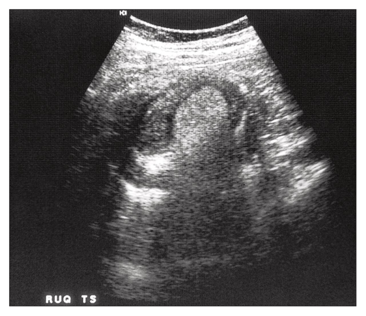
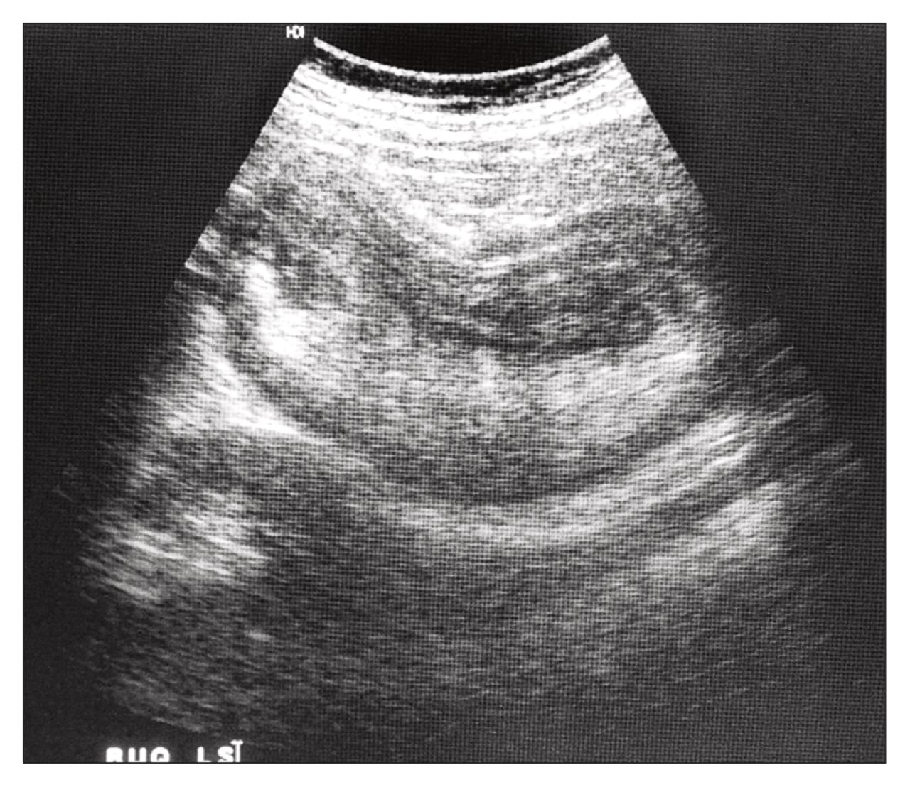
The classic sonographic features include the “dough-nut sign” in the transverse view (Fig. 1) and the “pseudokidney sign” in the longitudinal view (Fig. 2).3

FIG. 1
  • Download figure
  • Open in new tab
  • Download powerpoint
FIG. 1
FIG. 2
  • Download figure
  • Open in new tab
  • Download powerpoint
FIG. 2

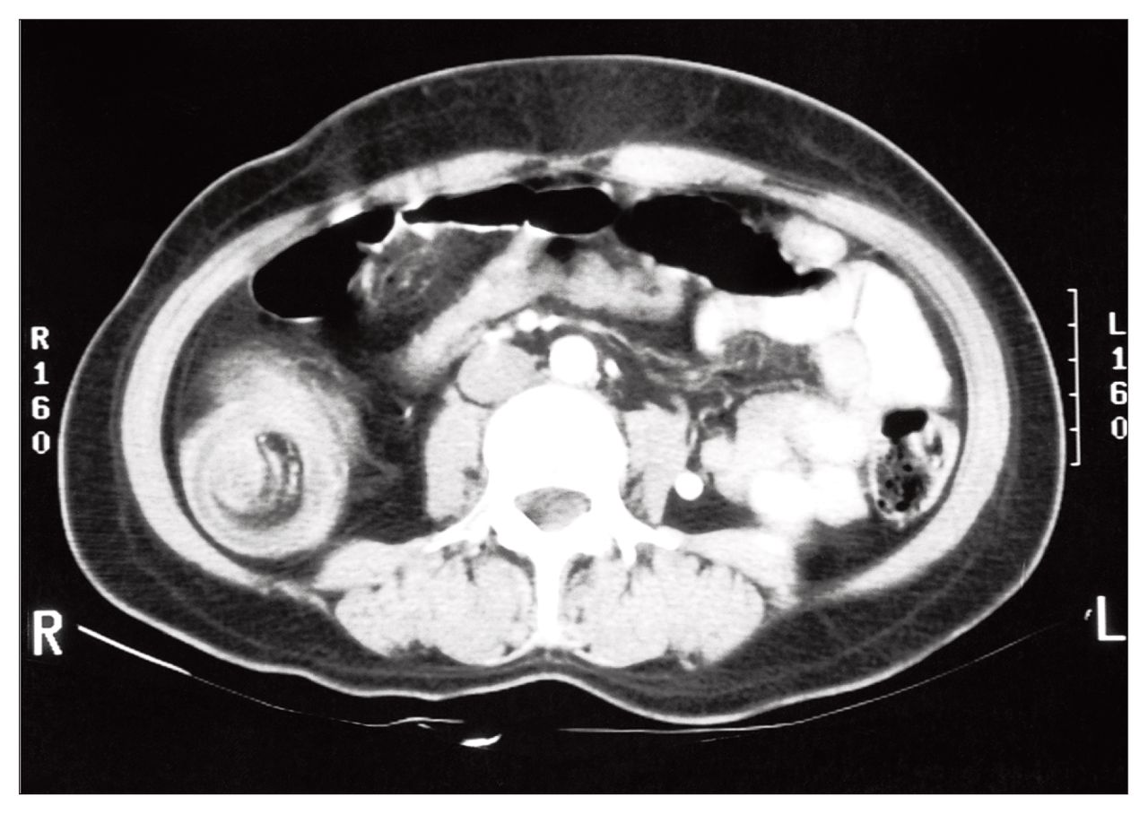
Abdominal CT reveals a mass lesion caused by a thickened segment of bowel due to telescoping of intussusceptum into the intussuscipiens and an eccentrically located low attenuated fatty mass that represents the invaginated mesentery (Fig. 3).4 In this case of ileocolic intussusception, CT at the level of the ileocecal valve revealed a tubular mass of mixed soft-tissue and fat projecting into the contrast-filled cecum (Fig. 4).

FIG. 3
  • Download figure
  • Open in new tab
  • Download powerpoint
FIG. 3
FIG. 4
  • Download figure
  • Open in new tab
  • Download powerpoint
FIG. 4

Laparotomy revealed a 13-cm segment of terminal ileum intussuscepting through the ileocecal valve into the ascending colon. The involved segment was resected. Histologic examination showed hemorrhagic necrosis of the terminal ileum but no other lesion. The patient recovered without complication.

References

  1. ↵
    1. Agha FP
    . Intussusception in adults. AJR 1986;146:527–31.
    OpenUrlCrossRefPubMed
  2. ↵
    1. Azar T,
    2. Berger DL
    . Adult intussusception. Ann Surg 1997;226(2):134–8.
    OpenUrlCrossRefPubMed
  3. ↵
    1. Boyle MJ,
    2. Arkell LJ,
    3. Williams JT
    . Ultrasonic diagnosis of adult intussusception [letter]. Am J Gastroenterol 1993;88: 617–8.
    OpenUrlPubMed
  4. ↵
    1. Donovan AT,
    2. Goldman SM
    . Computed tomography of ileocecal intussusception: mechanism and appearance. J Comput Assist Tomogr 1982;6:630–2.
    OpenUrlPubMed
PreviousNext
Back to top

In this issue

Canadian Journal of Surgery: 42 (5)
CAN J SURG
Vol. 42, Issue 5
1 Oct 1999
  • Table of Contents
  • Table of Contents (PDF)
  • Index by author

Article tools

Respond to this article
Print
Download PDF
Article Alerts
To sign up for email alerts or to access your current email alerts, enter your email address below:
Email Article

Thank you for your interest in spreading the word on CJS.

NOTE: We only request your email address so that the person you are recommending the page to knows that you wanted them to see it, and that it is not junk mail. We do not capture any email address.

Enter multiple addresses on separate lines or separate them with commas.
Soft-tissue case 29. diagnosis
(Your Name) has sent you a message from CJS
(Your Name) thought you would like to see the CJS web site.
CAPTCHA
This question is for testing whether or not you are a human visitor and to prevent automated spam submissions.
Citation Tools
Soft-tissue case 29. diagnosis
CAN J SURG Oct 1999, 42 (5) 344;

Citation Manager Formats

  • BibTeX
  • Bookends
  • EasyBib
  • EndNote (tagged)
  • EndNote 8 (xml)
  • Medlars
  • Mendeley
  • Papers
  • RefWorks Tagged
  • Ref Manager
  • RIS
  • Zotero
‍ Request Permissions
Share
Soft-tissue case 29. diagnosis
CAN J SURG Oct 1999, 42 (5) 344;
Digg logo Reddit logo Twitter logo Facebook logo Google logo Mendeley logo
  • Tweet Widget
  • Facebook Like

Related Articles

  • No related articles found.
  • Google Scholar

Cited By...

  • No citing articles found.
  • Google Scholar

Similar Articles

Content

  • Current issue
  • Past issues
  • Collections
  • Alerts
  • RSS

Authors & Reviewers

  • Overview for Authors
  • Publication Fees
  • Forms
  • Editorial Policies
  • Submit a manuscript

About

  • General Information
  • Staff
  • Editorial Board
  • Contact Us
  • Advertising
  • Reprints
  • Copyright and Permissions
  • Accessibility
  • CMA Civility Standards
CMAJ Group

Copyright 2023, CMA Impact Inc. or its licensors. All rights reserved. ISSN 2291-0026

All editorial matter in CJS represents the opinions of the authors and not necessarily those of the Canadian Medical Association or its subsidiaries.

To receive any of these resources in an accessible format, please contact us at CMAJ Group, 500-1410 Blair Towers Place, Ottawa ON, K1J 9B9; p: 1-888-855-2555; e: [email protected].

View CMA's Accessibility policy.

Powered by HighWire