


Abstract
Objectives: To establish the antioxidant status of rat intestinal tissues after ischemia–reperfusion and to determine if pretreatment with an allopurinol and antioxidant vitamin combination gives any protection against mucosal injury.
Experimental animals: Twenty rats were divided into 4 groups of 5 animals each.
Methods: Group 1 (control) rats were not subjected to ischemia–reperfusion and received no allopurinol plus vitamin combination; group 2 rats received vitamins C (200 mg/kg) and E (100 mg/kg) and allopurinol (50 mg/kg) combination daily for 3 days preoperatively; group 3 rats were subjected to ischemia–reperfusion only; and group 4 rats were subjected to ischemia–reperfusion and received the vitamin and allopurinol combination.
Main outcome measures: Activities of superoxide dismutase (SOD), glutathione peroxidase (GSH-Px) and catalase (CAT) enzymes, the level of thiobarbituric acid-reagent substances (TBARS) and histologic grading of tissue samples.
Results: SOD and GSH-Px activities were decreased, but the CAT activity and TBARS level increased. Pretreatment of the rats with the allopurinol-vitamin C-vitamin E combination did not have any significant effect on the enzyme activities. However, it resulted in important reductions in the TBARS tissue levels. Histologic investigation revealed significant mucosal injury in group 3 rats compared with group 4 rats (mean [and standard deviation] for grading, 4.6 [0.5] versus 1.8 [0.4]).
Conclusions: The enzymatic antioxidant defence system was significantly changed after ischemia–reperfusion and intestinal tissue was exposed to increased oxidant stress, the results of which were peroxidation of some cellular structures and increased concentrations of oxidative products. Although antioxidant treatment did not drastically affect the enzyme activities or afford complete protection of cellular structures against deformation, it apparently could eliminate oxygen radicals and prevent peroxidative reactions.
Hemorrhagic mucosal lesions are often found in the small intestine of patients during or after shock or hypertension.1 Mucosal injury is considered an important factor in the development of the shock state in intestinal tissues.2,3 The ischemic intestine appears especially sensitive to reperfusion injury, and the mechanism has been related to oxygen-derived free radicals.4–6 A cardiodepressive substance is thought to be released from ischemic intestine into the general circulation, and the mucosal damage initiates a cycle that leads to fatal circulatory deterioration. 2,3 Hypoxia of intestinal tissue during shock or hypotension plays an important role in the pathogenesis of the cellular injury.3,7 However, in local intestinal ischemia, additional tissue damage occurs when blood flow is restored. 6,8 There is general agreement that this additional tissue damage is caused by oxygen free radicals occurring during reoxygenation of the hypoxic tissue.6,8
It has been proposed that the basic source of the oxygen-derived free radicals is the xanthine oxidase (XO) system. 9,10 During ischemia, xanthine dehydrogenase is converted to XO. Although the mechanism of this conversion reaction is not understood in detail, protein kinase activation is thought to play a part.11 Accelerated breakdown of adenosine triphosphate (ATP) due to ischemia causes an accumulation of hypoxanthine and xanthine substrates for the XO enzyme. High substrate concentration and increased XO activity may both cause more superoxide radical production. It has been suggested that superoxide radicals produced by this reaction are responsible for the ischemia–reperfusion injury.4,8–10
We believe that increased radical concentrations may not be the only factor in this process. An inability to destroy the radicals produced may also play a role in the mucosal injury. As far as we know, no attempt has been made to elucidate this subject in the intestinal ischemia–reperfusion injury. We wished to investigate the enzymatic antioxidant potential of intestinal tissues exposed to ischemia–reperfusion injury and to establish whether allopurinol (an inhibitor of XO enzyme) plus antioxidant vitamin (vitamins C and E) pretreatment had any protective effect on the development of mucosal injury.
Materials and methods
Animals
In this study, 20 Sprague–Dawley rats each weighing about 220 g and about 3 months old were fed a standard laboratory diet. Ten of the rats were pretreated with a combination of allopurinol (50 mg/kg), vitamin E (100 mg/kg) and vitamin C (200 mg/kg) daily for 3 days before operation. Allopurinol was given orally, and vitamins were given by intramuscular injection. Animals were divided into 4 groups of 5 as follows: group 1 — control (no ischemia–reperfusion, no allopurinol plus vitamin combination); group 2 — allopurinol plus vitamin combination only; group 3 — ischemia–reperfusion only; group 4 — ischemia–reperfusion and allopurinol plus vitamin combination. All rats were first anesthetized with an intramuscular injection of detamine hydrochloride (80 mg/kg). Intestinal ischemia was produced in groups 3 and 4 by clamping the superior mesenteric artery for 40 minutes; this was followed by 40 minutes of reperfusion. After the ischemia–reperfusion process, intestinal tissue samples were obtained from the jejunum; some were used for histologic investigation, others were frozen at −30 °C for about 3 days to be used in the biochemical assays.
Histologic examination
After fixation in 5% formalin, samples of intestinal tissue were embedded in paraffin, cut at about 5 μm and stained with hematoxylin–eosin. The slides were coded and examined for morphologic changes by light microscopy. Samples were graded according to the scale of Chiu and associates12 as follows: 0 = normal villi, 1 = subepithelial space at the tip of the villus, 2 = moderate separation of mucosa from the lamina propria, 3 = extensive subepithelial separation of mucosa from the lamina propria down the sides of the villus and ulceration of the villus tip, 4 = denuded villi, 5 = disintegration of the lamina propria, hemorrhagic ulceration.
Biochemical assays
Parts of the tissue samples were homogenized in physiologic saline solution and centrifuged at 5500 × g for 30 minutes. Clear supernatant was taken for the enzymatic assays. Superoxide dismutase (SOD) (measured in U/mg protein) and glutathione peroxidase (GSH-Px) and catalase (CAT) (measured in IU/mg protein) activities were determined as previously described. 13–15 The protein content was determined by the method of Lowry and associates.16 One unit of SOD activity was defined as the amount of enzyme protein causing 50% inhibition in the nitroblue tetrazolium reduction rate. Other parts of the samples were used to determine levels of thio bar bituric acid-reagent substances (TBARS).17
Statistical analysis
The results were analysed by Tukey’s test and by analysis of variance. A p value of less than 0.05 was considered significant.
Results
The mean (and standard deviation) values of the enzyme activities and TBARS levels in the intestinal tissues of the rats are shown in Table I. SOD and GSH-Px activities were decreased, but the activity of CAT increased in the intestinal tissues after ischemia–reperfusion. Levels of TBARS were higher in the ischemic tissues than in the tissues of control and antioxidant-treated rats. Although antioxidant pretreatment did not have a significant effect on the enzyme activities, it lowered tissue levels of TBARS.
Mean (and SD) Values for Superoxide Dismutase (SOD), Glutathione Peroxidase (GSH-Px) and Catalase (CAT) Enzyme Activities and Thiobarbituric Acid-Reagent Substances (TBARS) Levels in the Intestinal Tissues of Rats After Ischemia–Reperfusion With or Without Pretreatment With Vitamins and Allopurinol
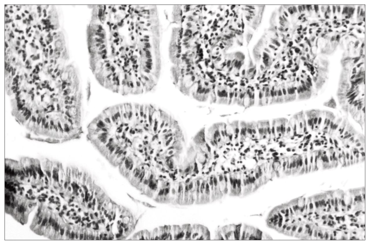
Histologic examination showed serious mucosal injury in group 3 and mod erate mucosal injury in group 4. There was no difference histologically between groups 1 and 2 (Table II, Figs. 1 to ⇓3).

A tissue sample from group 1 (control), showing normal intestinal villi (hematoxylin–eosin, original magnification × 40).
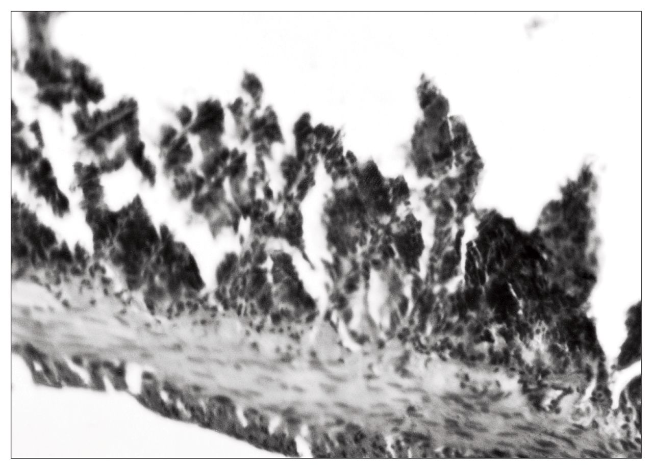
A tissue sample from group 3 (ischemia–reperfusion) shows disintegration of the lamina propria, ulceration of villus tips and denuded villi (hematoxylin–eosin, original magnification × 40).

A tissue sample from group 4 (ischemia–reperfusion and pretreatment with vitamins C and E and allopurinol) shows separation of mucosa from the lamina propria (hematoxylin–eosin, original magnification × 40).
Histopathological Grading of Rat Intestinal Tissues in the 4 Groups
Discussion
Hemorrhagic mucosal lesions of the small intestine are often seen in experimental animals subjected to shock protocols. 3,17,18 Similar lesions were also seen histologically in this study. In this respect, our results confirm the earlier observations that significant mucosal damage occurs after ischemia–reperfusion and that this damage is caused by oxygen-derived free radicals. Free radicals can cause peroxidation in lipid components of cellular membranes, resulting in increased levels of TBARS in the tissues affected.5,19,20
In previous studies, the main problem in ischemia–reperfusion injury was thought to be the overproduction of oxygen radicals due to accelerated XO reactions.4,8,21,22 In this study, we also found that the enzymatic antioxidant defence system was meaningfully changed in the intestinal tissues after ischemia–reperfusion. Cytoplasmic components of the enzymatic antioxidant defence system, namely SOD and GSH-Px, were suppressed, and lysosomal CAT was activated. Oxidant stress is known to upregulate gene expression of antioxidant enzymes and may be mediated by downregulation of antioxidant gene expression. For these, various time periods have been reported. For example, in a previous study, manganous SOD gene expression was reported to be induced within 1 hour of treatment,23 and in another study, transcript levels of CAT, manganous SOD and copper/ zinc SOD were reported to be down-regulated in 8 hours.24
In the ischemia–reperfusion process it seems that on one hand the production of oxygen radicals is increased and on the other hand their normal catabolism is prevented because of reduced cytoplasmic antioxidant capacity, the results of which are accelerated peroxidation reactions.
Although CAT activity was increased, we believe this was of secondary importance since this CAT enzyme was mainly localized in cell lysosomes. In fact, this activation may be a compensatory mechanism against lowered GSH-Px activity because both enzymes use hydrogen peroxide as substrate, the product of SOD-catalyzed dismutation reaction.
Whatever the reasons were, the result was that the cytoplasmic antioxidant defence system of intestinal tissue was reduced after ischemia–reperfusion and could not eliminate oxygen radicals produced at a normal catabolic rate. Thus, cellular peroxidation reactions were accelerated, resulting in mucosal damage. Our results also show that these consecutive events can be prevented in part by a combination of vitamins C and E and allopurinol. In a previous study, ascorbic acid was also shown to prevent peroxidative tissue injury.25 Allopurinol has dual functions: as an XO inhibitor and an antioxidant.26 Pretreatment of the rats with these antioxidants effectively prevents superoxide radical production by the XO enzymatic system and eliminates free radicals produced by other sources. This is clearly seen from decreased level of TBARS as a result of antioxidant pretreatment. Histologic examination also indicated that antioxidant pretreatment did not afford complete protection of cellular structures against ischemic deformation although it pre vented oxidative reactions. This means that increased free radical production is not the only factor in the development of ischemic damage and that other radical-independent factors play a role.17,27
Conclusions
Intestinal mucosa is subjected to oxidant stress because of an overproduction of free radicals and an inability to destroy them, during and after ischemia–reperfusion. Allopurinol plus antioxidant pretreatment eliminates the oxidant stress and protects intestinal mucosal tissue against free radicals.
- Accepted December 15, 1998.
References
In this issue

Article tools


Related Articles
Cited By...
- No citing articles found.