
Main menu

  • Home
  • Content
    • Current issue
    • Past issues
    • Sections
    • Collections
  • Podcasts
  • Author Info
    • Overview for authors
    • Publication fees
    • Forms
    • Editorial policies
    • Submit a manuscript
    • Open access
  • Careers
  • Alerts
    • Email alerts
    • RSS
  • About
    • General information
    • Staff
    • Editorial Board
    • Contact
  • CMAJ JOURNALS
    • CMAJ
    • CMAJ Open
    • JAMC
    • JPN

User menu

Search

  • Advanced search
CJS
  • CMAJ JOURNALS
    • CMAJ
    • CMAJ Open
    • JAMC
    • JPN
CJS

Advanced Search

  • Home
  • Content
    • Current issue
    • Past issues
    • Sections
    • Collections
  • Podcasts
  • Author Info
    • Overview for authors
    • Publication fees
    • Forms
    • Editorial policies
    • Submit a manuscript
    • Open access
  • Careers
  • Alerts
    • Email alerts
    • RSS
  • About
    • General information
    • Staff
    • Editorial Board
    • Contact
  • Subscribe to our alerts
  • RSS feeds
  • Follow CJS on Twitter
Radiology for the Surgeon

Musculoskeletal case 7. Diagnosis

CAN J SURG December 01, 1999 42 (6) 432;
  • Article
  • Figures & Tables
  • Responses
  • Metrics
  • PDF
Loading

Large femoral geode associated with osteoarthritis of the hip joint

The plain radiograph of the right hip (Fig. 1) demonstrated a large lytic lesion in the proximal femur, which on magnetic resonance imaging (Fig. 2) was shown to be cystic and consistent with a large subarticular cyst. The large femoral geode was confirmed at right total hip arthroplasty.

FIG. 1
  • Download figure
  • Open in new tab
  • Download powerpoint
FIG. 1

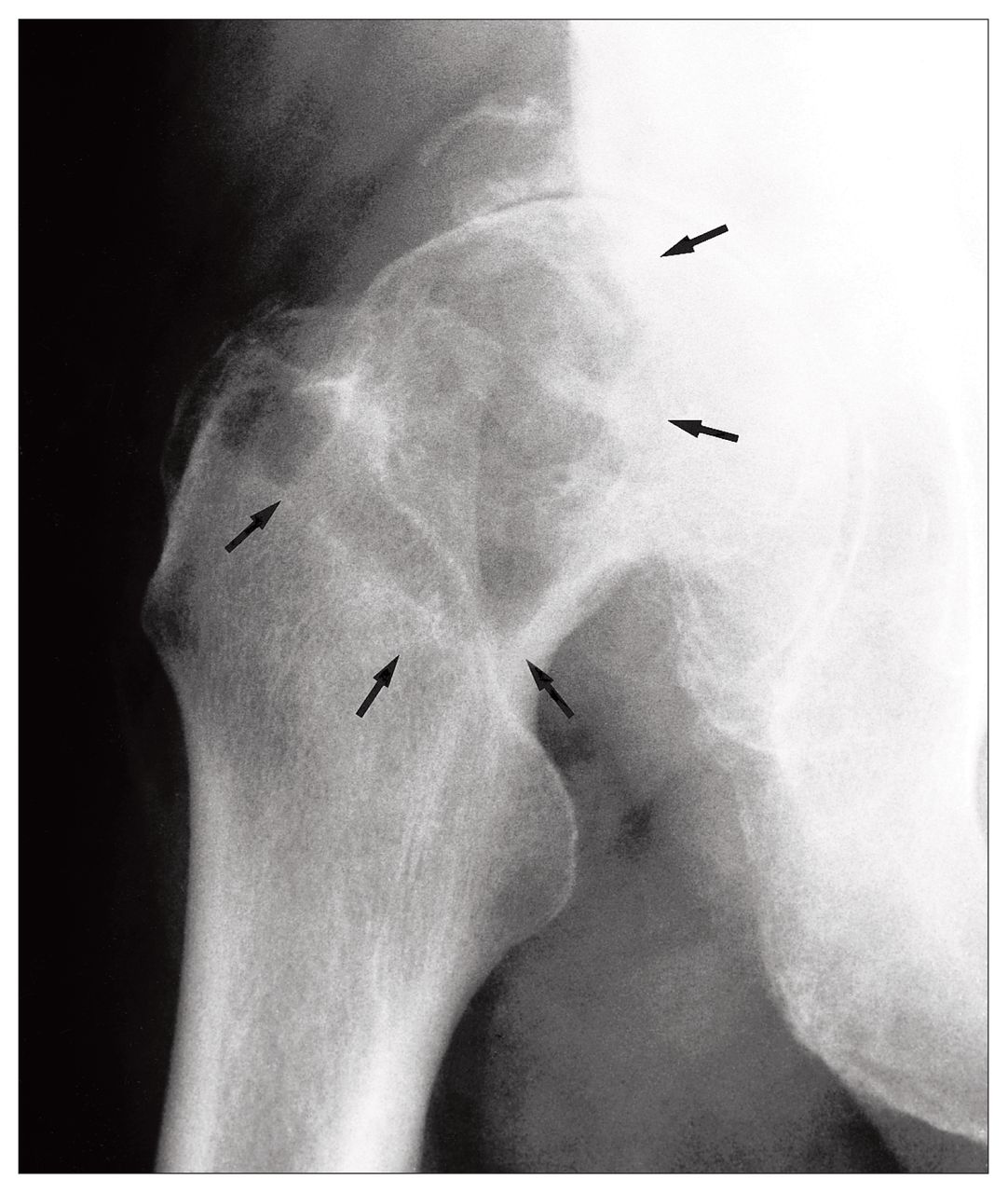
Anteroposterior radiograph of the right hip reveals a large well-defined lytic lesion in the lateral aspect of the proximal femoral meta-piphysis and epiphysis (arrows). Extensive degenerative changes are present with joint space loss, osteophytes and subchondral sclerosis.

FIG. 2
  • Download figure
  • Open in new tab
  • Download powerpoint
FIG. 2

Coronal spin-echo, T1-weighted (left) and T2-weighted (right) images of the right hip show the lesion having well-defined borders with low signal on T1 and high signal on T2 consistent with fluid. The lesion did not enhance after administration of contrast medium. The extensive degenerative changes are shown.

The term geode is used in geology to describe small hollow rocks lined with crystals.1 Subchondral cysts, or geodes, are common findings in degenerative joint disease. They can be seen in rheumatoid arthritis, osteoarthritis, calcium pyrophosphate deposition disease and avascular necrosis.2 The cysts typically form within the weight-bearing portion of subchondral bone. Pathologically, they appear between thickened trabeculae and are of variable size and multiplicity. The pathogenesis is thought to be due either to synovial fluid intrusion or to bony contusion with subsequent fracture and vascular compromise of subchondral bone, causing cystic necrosis.1

A geode can mimic a more ominous process such as chondroblastoma, giant cell tumour and skeletal metastasis, especially when it occurs in the presence of a normal-appearing joint.2 A femoral neck geode without joint involvement in a rheumatoid patient has been reported.3

The MRI appearance of the geode in this case is typical. With the use of appropriate imaging techniques, biopsy of such a lesion can be avoided.2,3

References

  1. ↵
    1. Resnick D
    . Diagnosis of bone and joint disorders. Philadelphia: W.B. Saunders; 1988. p. 1375–81.
  2. ↵
    1. Helms CA
    . Fundamentals of skeletal radiology. 2nd ed. Toronto: W.B. Saunders; 1995. pp. 30, 59–61, 119–20.
  3. ↵
    1. Morrey BF
    . Rheumatoid pseudocyst (geode) of the femoral neck without apparent joint involvement. Mayo Clin Proc 1987;62(5):407–11.
    OpenUrlPubMed
PreviousNext
Back to top

In this issue

Canadian Journal of Surgery: 42 (6)
CAN J SURG
Vol. 42, Issue 6
1 Dec 1999
  • Table of Contents
  • Table of Contents (PDF)
  • Index by author

Article tools

Respond to this article
Print
Download PDF
Article Alerts
To sign up for email alerts or to access your current email alerts, enter your email address below:
Email Article

Thank you for your interest in spreading the word on CJS.

NOTE: We only request your email address so that the person you are recommending the page to knows that you wanted them to see it, and that it is not junk mail. We do not capture any email address.

Enter multiple addresses on separate lines or separate them with commas.
Musculoskeletal case 7. Diagnosis
(Your Name) has sent you a message from CJS
(Your Name) thought you would like to see the CJS web site.
CAPTCHA
This question is for testing whether or not you are a human visitor and to prevent automated spam submissions.
Citation Tools
Musculoskeletal case 7. Diagnosis
CAN J SURG Dec 1999, 42 (6) 432;

Citation Manager Formats

  • BibTeX
  • Bookends
  • EasyBib
  • EndNote (tagged)
  • EndNote 8 (xml)
  • Medlars
  • Mendeley
  • Papers
  • RefWorks Tagged
  • Ref Manager
  • RIS
  • Zotero
‍ Request Permissions
Share
Musculoskeletal case 7. Diagnosis
CAN J SURG Dec 1999, 42 (6) 432;
Digg logo Reddit logo Twitter logo Facebook logo Google logo Mendeley logo
  • Tweet Widget
  • Facebook Like

Related Articles

  • No related articles found.
  • Google Scholar

Cited By...

  • No citing articles found.
  • Google Scholar

Similar Articles

Content

  • Current issue
  • Past issues
  • Collections
  • Alerts
  • RSS

Authors & Reviewers

  • Overview for Authors
  • Publication Fees
  • Forms
  • Editorial Policies
  • Submit a manuscript

About

  • General Information
  • Staff
  • Editorial Board
  • Contact Us
  • Advertising
  • Reprints
  • Copyright and Permissions
  • Accessibility
  • CMA Civility Standards
CMAJ Group

Copyright 2023, CMA Impact Inc. or its licensors. All rights reserved. ISSN 2291-0026

All editorial matter in CJS represents the opinions of the authors and not necessarily those of the Canadian Medical Association or its subsidiaries.

To receive any of these resources in an accessible format, please contact us at CMAJ Group, 500-1410 Blair Towers Place, Ottawa ON, K1J 9B9; p: 1-888-855-2555; e: [email protected].

View CMA's Accessibility policy.

Powered by HighWire