
Main menu

  • Home
  • Content
    • Current issue
    • Past issues
    • Sections
    • Collections
  • Podcasts
  • Author Info
    • Overview for authors
    • Publication fees
    • Forms
    • Editorial policies
    • Submit a manuscript
    • Open access
  • Careers
  • Alerts
    • Email alerts
    • RSS
  • About
    • General information
    • Staff
    • Editorial Board
    • Contact
  • CMAJ JOURNALS
    • CMAJ
    • CMAJ Open
    • JAMC
    • JPN

User menu

Search

  • Advanced search
CJS
  • CMAJ JOURNALS
    • CMAJ
    • CMAJ Open
    • JAMC
    • JPN
CJS

Advanced Search

  • Home
  • Content
    • Current issue
    • Past issues
    • Sections
    • Collections
  • Podcasts
  • Author Info
    • Overview for authors
    • Publication fees
    • Forms
    • Editorial policies
    • Submit a manuscript
    • Open access
  • Careers
  • Alerts
    • Email alerts
    • RSS
  • About
    • General information
    • Staff
    • Editorial Board
    • Contact
  • Subscribe to our alerts
  • RSS feeds
  • Follow CJS on Twitter
Original Article

Value of DNA ploidy and S-phase fraction as prognostic factors in stage III cutaneous melanoma

Ginette Martin, Fawaz Halwani, Henry Shibata and Sarkis Meterissian
CAN J SURG February 01, 2000 43 (1) 29-34;
Ginette Martin
*Department of Surgical Oncology, Royal Victoria Hospital, McGill University, Montreal, Que.
  • Find this author on Google Scholar
  • Find this author on PubMed
  • Search for this author on this site
Fawaz Halwani
†Department of Pathology, Royal Victoria Hospital, McGill University, Montreal, Que.
  • Find this author on Google Scholar
  • Find this author on PubMed
  • Search for this author on this site
Henry Shibata
*Department of Surgical Oncology, Royal Victoria Hospital, McGill University, Montreal, Que.
  • Find this author on Google Scholar
  • Find this author on PubMed
  • Search for this author on this site
Sarkis Meterissian
*Department of Surgical Oncology, Royal Victoria Hospital, McGill University, Montreal, Que.
  • Find this author on Google Scholar
  • Find this author on PubMed
  • Search for this author on this site
  • Article
  • Figures & Tables
  • Responses
  • Metrics
  • PDF
Loading

Abstract

Objective: To determine the prognostic value of flow cytometric analysis (S-phase fraction and DNA index) performed on lymph-node metastases of patients with stage III melanoma.

Design: A retrospective chart review with flow cytometric analysis of paraffin-embedded tissues.

Setting: A university teaching hospital.

Patients: Among 332 patients with cutaneous melanoma, 33 with stage III were identified. Distant metastases developed in 16 patients; 17 had no further recurrence. Charts were reviewed to obtain clinicopathologic parameters such as sex, age, location of the primary tumour, histologic features, presence or absence of ulceration, and Clark’s and Breslow’s levels.

Intervention: DNA ploidy and S-phase fraction were determined on the paraffin-embedded nodes.

Main outcome measures: The groups with or without recurrence were compared in terms of disease-free survival (DFS) and overall survival (OS). These survival parameters were correlated with DNA ploidy and S-phase fraction.

Results: By univariate analysis, clinicopathologic factors did not predict OS. A higher Clark’s level of invasion and more than 3 positive lymph nodes were associated with shorter DFS (p < 0.05). Tumour thickness and S-phase fraction did not correlate with either DFS or OS. Patients with diploid lymph-node metastases had an 87% 12-month survival compared with 41% for those with aneuploid tumours.

Conclusions: DNA ploidy may be used as a prognostic index in patients with lymph-node metastases. This could be particularly useful in the context of sentinel lymph-node mapping by which more patients are being identified with single microscopic lymph-node involvement.

The incidence of cutaneous malignant melanoma is increasing at a rate faster than any other cancer in United States and Canada. According to the American Cancer Society, approximately 7200 patients will die of metastatic melanoma over the next 10 years.1 The prognosis in patients with stages I and II cutaneous melanoma can be predicted using a number of morphologic variables including Breslow’s thickness, Clark’s level of invasion, the presence of ulceration or regression, host inflammatory cell response, growth pattern and blood vessel invasion. 2–8 These have been well characterized, and multivariate analysis has identified Breslow’s thickness as the single most important prognostic factor in primary cutaneous malignant melanoma. For stage III melanoma (TNM classification, regional nodal involvement), however, the number of prognostic factors is far less and includes only number and percentage of positive lymph nodes as well as the presence of extranodal extension.9–12 With the advent of sentinel lymph-node mapping, increasing numbers of patients are being identified with single microscopic nodal involvement, making prediction of the prognosis more difficult.13,14

To further classify patients into high- and low-risk groups, flow cytometry has been used primarily in stages I and II melanoma, with DNA ploidy emerging as an important prognostic factor.15–20 Little work has been done on stage III melanoma. A single study has demonstrated that DNA analysis of melanoma metastases with flow cytometry might provide additional prognostic information.21

Recent publications have shown benefits of interferon-alpha therapy in patients with high-risk resected cutaneous melanoma.21–23 The purpose of this study was to determine whether DNA ploidy and S-phase fraction of lymph-node metastases could improve prognostication in patients having stage III melanoma, thus providing a better guide to the use of adjuvant therapy.

Patients and methods

From 1986 to 1996, 332 patients with primary cutaneous melanoma were seen at the Royal Victoria Hospital in Montreal. Of these, 33 patients (21 men, 12 women) underwent a therapeutic lymph-node dissection with paraffin-embedded lymph-node specimens available for analysis. DNA analysis was carried out on all specimens, but determination of the S-phase fraction was possible on only 30 (91%). The median age of the 33 patients was 54.2 years. Medical charts were reviewed for clinicopathological information such as age, sex, anatomic location of the primary lesion, Breslow’s thickness, Clark’s level of invasion, presence of ulceration, number of positive lymph nodes and extranodal extension. Patient survival was determined as the time from nodal dissection to the date of last follow-up. Among these 33 patients, 5 had radiotherapy, 2 received immunotherapy and 1 had chemotherapy. Follow-up was completed at the time of data collection and all deaths were attributable to melanoma. The median follow-up was 29 months.

Flow cytometry

Tissue sections (50 μm thick) from representative formalin-fixed paraffin blocks of metastatic lymph nodes were taken. Specimens were dewaxed in xylene and rehydrated in a stepwise fashion. Enzymatic digestion was done using pepsin solution (0.5%; Sigma Chemical, St. Louis). After a wash in phosphate-buffered saline, the suspended nuclei were stained with propidium iodide and incubated in the dark at 4 °C overnight. The nuclear suspension was then filtered and submitted to flow cytometric analysis using an argon laser at 488 nm (Epics Profile II; Coulter Electronics, Hialeah, Fla.). Specimens were considered diploid if only one G0/G1 peak was present. Aneuploid histograms were defined as the presence of at least 2 separate peaks, each having at least 5% of the total number of events and each with a coefficient of variance of less than 5 (Fig. 1). The percentage of cells in the S-phase of the cell cycle was determined using the multicycle software with a nonlinear least square statistic and debris subtraction (Phoenix Flow System, San Diego).

FIG. 1
  • Download figure
  • Open in new tab
  • Download powerpoint
FIG. 1

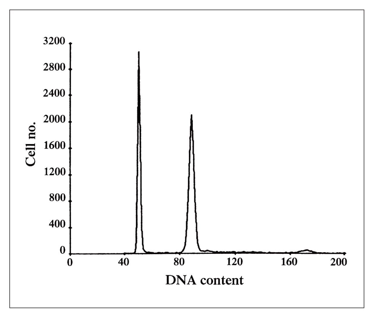
DNA ploidy histogram of a melanoma metastatic to an inguinal lymph node. The first peak at channel 50 represents the diploid content of the normal lymphoid tissue, serving as an internal control. The second peak at channel 97 represents the aneuploid melanoma cells with a DNA index of 2. The small peak on the right of the histogram is the dividing population of the malignant cells in the G2/M phase of the cell cycle.

Statistical analysis

Survival analysis was performed using the Kaplan–Meier method. The Mantel–Cox statistical test was used for all DNA analyses and univariate analysis for the clinocopathological characteristics.

Results

The median (and standard deviation) disease-free survival (DFS) for the entire group was 10.3 (7.6) months, and median overall survival (OS) was 14.5 (6.3) months. The distribution and outcome of the 33 patients according to their clinicopathological characteristics are shown in Table I. Among the various factors analysed, only sex approached significance for predicting overall survival (p = 0.068). Clark’s level of invasion appeared to be a significant predictor for DFS (p = 0.03); however, there was only 1 patient in the level V group.

View this table:
  • View inline
  • View popup
Table I

Survival and Clinicopathologic Features of 33 Patients Having Stage III Cutaneous Melanoma

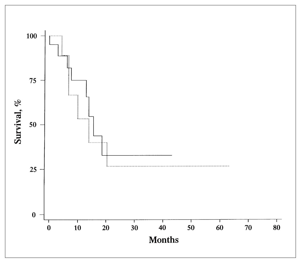
Analysis of lymph-node-related variables is summarized in Table II. Only DNA index (DI) correlated with outcome. The 12-month survival was only 41% for those with aneuploid lymph nodes (DI different from 1), compared with 87% for those with a DI equal to 1 (p = 0.06) (Fig. 2). An S-phase fraction lower than 6% was also associated with higher survival at 12 months (70% compared with 53% for an S-phase fraction 6% or greater). This difference, however, was not statistically significant (Fig. 3).

FIG. 2
  • Download figure
  • Open in new tab
  • Download powerpoint
FIG. 2

Kaplan–Meier analysis of 12-month survival of patients with stage III melanoma according to ploidy status. Patients with aneuploid lymph nodes had an index different from 1 (dotted line); those with diploid lymph nodes had an index equal to 1 (solid line) (p = 0.06).

FIG. 3
  • Download figure
  • Open in new tab
  • Download powerpoint
FIG. 3

Kaplan–Meier analysis of 12-month survival of patients with stage III melanoma according to low (less than 6, solid line) and high (6 or greater, dotted line) S-phase fractions (p = 0.71).

View this table:
  • View inline
  • View popup
Table II

Survival and Flow Cytometry Results in 33 Patients Having Stage III Cutaneous Melanoma

Discussion

Although cutaneous malignant melanoma represents less than 2% of all cancers,1 its incidence has steadily increased over the last 2 decades. Primary or nonmetastatic melanoma has been studied extensively, with a number of clinicopathological parameters being identified as important predictors of prognosis. These include Breslow’s thickness, Clark’s level of invasion, presence or absence of ulceration and tumour location.2–8 Unfortunately, none can be used to predict prognosis of stage III patients. In fact, in this group, studies have shown that only the number of lymph nodes involved and presence of extracapsular extension are significant predictors of outcome.9–12

Two recent developments have generated a need to subdivide stage III patients into high- and low-risk groups. One is the advent of sentinel lymph-node mapping, which allows the clinician to identify an involved node without performing a complete node dissection with its attendant morbidity. Morton and colleagues24 have shown that when the sentinel lymph node is invaded, at least 78% of patients have only one lymph node involved in the entire node basin. Thus, we are beginning to identify a large cohort of patients with stage III disease in whom traditional prognostic factors are no longer useful. Second, Kirkwood and associates23 have recently demonstrated the efficacy of interferon alpha in stage III melanoma. As we begin using this agent clinically, it will become imperative that we develop novel prognostic markers to predict subsets of patients who will respond best. The purpose of our study was to determine whether DNA ploidy or S-phase fraction can be used to predict DFS and OS in patients with stage III melanoma.

S-phase fraction was found not to be related to OS or DFS. Hansson and colleagues25 (Table III16,17,19,20,22,25–30) found that the S-phase fraction was related to survival; however, they used 10% as their cut-off compared with the more stringent 6% used in this study. In fact only 2 of our patients had an S-phase fraction greater than 10%. Karlsson and associates,26 using paraffinembedded specimens, demonstrated that the S-phase fraction is a significant predictor of survival. In our study, the S-phase fraction was higher in those patients who died, but this difference did not reach statistical significance; therefore larger studies should be performed to obtain an adequate number of patients before reaching a conclusion about the usefulness of this parameter.

View this table:
  • View inline
  • View popup
Table III

Review of the Literature on Melanoma and Flow Cytometry

In this study, DNA ploidy measured on metastatic lymph nodes was associated with improved survival with a probability value approaching statistical significance. Heaton and associates, 22 also looking at metastatic lymph nodes, found that DNA indices greater than 2 were significant predictors of poor patient survival. Hansson and colleagues25 found a similar correlation between prognosis and DNA index. Only Muhonen and colleagues,27 surprisingly, found that aneuploidy was associated with longer survival. This unusual association may be explained by the fact that most of their patients received systemic chemotherapy, and aneuploid tumours seem to have a greater response to chemotherapy than do diploid tumours. Most other published papers on DNA ploidy examined stages I and II disease, and the primary cutaneous lesions rather than the metastases as in our study.

With the advent of sentinel lymph-node mapping and the increasing use of interferon alpha we believe that accurate prognostic markers must be identified. This study has investigated one potential marker technique, flow cytometry. Results must be confirmed on a larger cohort of patients, but it appears that DNA ploidy may be useful for better stratification and further subset analysis of patients assigned to adjuvant therapy.

Footnotes

  • Presented in abstract form at the meeting of the American Association for Cancer Research, Washington, DC, Apr. 20 to 24, 1997.

  • Accepted December 10, 1998.

References

  1. ↵
    1. Avril MF,
    2. Cascinelli N,
    3. Cristofolini M
    . Clinical diagnosis of melanoma. Geneva: WHO Melanoma Program Publications; 1994. No. 3.
  2. ↵
    1. Eldh J,
    2. Boeryd B,
    3. Peterson LE
    . Prognostic factors in cutaneous malignant melanoma in stage I. A clinical, morphological and multivariate analysis. Scand J Plast Reconstr Surg 1978; 12(3):243–55.
    OpenUrlPubMed
    1. Day CL Jr.,
    2. Lew RA,
    3. Mihm MC Jr.,
    4. Sober AJ,
    5. Harris MN,
    6. Kopf AW,
    7. et al
    . A multivariate analysis of prognostic factors for melanoma patients with lesions 3.65 mm in thickness. Ann Surg 1982;195:44–9.
    OpenUrlCrossRefPubMed
    1. Balch CM,
    2. Wilkerson JA,
    3. Murad TM,
    4. Soong S-J,
    5. Ingalls AL,
    6. Maddox WA
    . The prognostic significance of ulceration of cutaneous melanoma. Cancer 1980;45:3012–7.
    OpenUrlCrossRefPubMed
    1. Day CL Jr.,
    2. Mihm MC,
    3. Sober AJ,
    4. Harris MN,
    5. Kopf AW,
    6. Fitzpatrick TS,
    7. et al
    . Prognostic factors for melanoma patients with lesions 0.76–1.69 mm in thickness. Ann Surg 1982;195:30–4.
    OpenUrlPubMed
    1. Balch CM,
    2. Houghton AN,
    3. Milton GW,
    4. Sober AJ,
    5. Soong S-J
    , editors. Cutaneous melanoma. 2nd ed. New York: J.B. Lippincott; 1992. p. 188–99.
    1. Balch CM,
    2. Houghton AN,
    3. Milton GW,
    4. Sober AJ,
    5. Soong S-J
    , editors. Cutaneous melanoma. 2nd ed. New York: J.B. Lippincott; 1992. p. 165–87.
  3. ↵
    1. Balch CM,
    2. Murad TM,
    3. Soong S-J,
    4. Ingallo AL,
    5. Halpern NB,
    6. Maddox WA
    . A multifactorial analysis of melanoma: prognostic histopathological features comparing Clark’s and Breslow’s staging methods. Ann Surg 1978;188: 732–42.
    OpenUrlPubMed
  4. ↵
    1. Balch CM,
    2. Soong SJ,
    3. Murad TM,
    4. Ingalls AL,
    5. Maddox WA
    . A multifactorial analysis of melanoma: III. Prognostic factors of melanoma patients with lymph node metastases (stage II). Ann Surg 1981;193(3):377–88.
    OpenUrlCrossRefPubMed
    1. Bevilacqua RG,
    2. Coit DG,
    3. Rogatko A,
    4. Younes RN,
    5. Brennan MF
    . Axillary dissection in melanoma. Prognostic variables in node-positive patients. Ann Surg 1990;212:125–31.
    OpenUrlPubMed
    1. Callery C,
    2. Cochran A,
    3. Roe DJ,
    4. Rees N,
    5. Nathanson SD,
    6. Benedetti JK,
    7. et al
    . Factors prognostic for survival in patients with malignant melanoma spread to regional lymph nodes. Ann Surg 1982;196:69–75.
    OpenUrlPubMed
  5. ↵
    1. Coit D,
    2. Rogatki A,
    3. Brennan M
    . Prognostic factors in patients with melanoma metastatic to axillary or inguinal lymph nodes. A multivariate analysis. Ann Surg 1991;214:627–36.
    OpenUrlCrossRefPubMed
  6. ↵
    1. Morton DL,
    2. Wen DR,
    3. Wong JH,
    4. Economou JS,
    5. Cagle LA,
    6. Storn FK,
    7. et al
    . Technical details of intraoperative lymphatic mapping for early stage melanoma. Arch Surg 1992;127:392–9.
    OpenUrlCrossRefPubMed
  7. ↵
    1. Krag DN,
    2. Meijer SJ,
    3. Weaver PL,
    4. Loggie BW,
    5. Harlow SP,
    6. Tanabe KK,
    7. et al
    . Minimal access surgery for staging of malignant melanoma. Arch Surg 1995; 130:654–60.
    OpenUrlCrossRefPubMed
  8. ↵
    1. Lindholm C,
    2. Hofer PA,
    3. Jonsson H,
    4. Tribukait B
    . Flow DNA cytometric findings of paraffin-embedded primary cutaneous melanoms related to prognosis. Virchows Arch B Cell Pathol 1989; 58:147–51.
    OpenUrlPubMed
  9. ↵
    1. Lindholm C,
    2. Hofer PA,
    3. Jonsson H
    . Single cell DNA cytophotometry in clinical stage I malignant melanoma. Acta Oncol 1990;29:147–50.
    OpenUrlCrossRefPubMed
  10. ↵
    1. Kheir SM,
    2. Benes SD,
    3. von Roenn JH,
    4. Soong S-J,
    5. Coon JS
    . Prognostic significance of DNA aneuploidy in stage I cutaneous melanoma. Ann Surg 1988; 207:455–61.
    OpenUrlPubMed
    1. Gattuso P,
    2. Reddy V,
    3. Solans E,
    4. Kathuria S,
    5. Aranha GV,
    6. Jacobs HK,
    7. et al
    . Is DNA ploidy of prognostic significance in stage I cutaneous melanoma? Surgery 1990;108:702–9.
    OpenUrlPubMed
  11. ↵
    1. Sondergaard K,
    2. Larsen JK,
    3. Moller U,
    4. Christensen IJ,
    5. Hou-Jensen K
    . DNA ploidy characteristics of human malignant melanoma analysed by flow cytometry and compared with histology and clinical course. Virchows Arch B Cell Pathol Incl Mol Pathol 1983; 42:43–52.
    OpenUrlPubMed
  12. ↵
    1. von Roenn JH,
    2. Kheir SM,
    3. Wolter JM,
    4. Coon JS
    . Significance of DNA abnormalities in primary malignant melanoma and nevi: a retrospective flow cytometric study. Cancer Res 1986;46:3192–5.
    OpenUrlAbstract/FREE Full Text
  13. ↵
    1. Balch CM,
    2. Buzaid AC
    . Finally a successful adjuvant therapy for high-risk melanoma. J Clin Oncol 1996;14:1–3.
    OpenUrlPubMed
  14. ↵
    1. Heaton KM,
    2. Rippon MB,
    3. El-Naggar A,
    4. Tucker SL,
    5. Ross MI,
    6. Balch CM
    . Prognostic implications of DNA index in patients with stage III cutaneous melanoma. Am J Surg 1993; 166: 648–53.
    OpenUrlPubMed
  15. ↵
    1. Kirkwood JM,
    2. Strawderman MH,
    3. Ernstoff MS,
    4. Smith TJ,
    5. Borden EC,
    6. Blum RH
    . Interferon alfa-2b adjuvant therapy of high-risk resected cutaneous melanoma: the Eastern Cooperative Oncology Group Trial EST 1684. J Clin Oncol 1996;14(1):7–17.
    OpenUrlAbstract
  16. ↵
    1. Morton DL,
    2. Wen DR,
    3. Cochran AV
    . Management of early stage melanoma by intraoperative lymphatic mapping and selective lymphadectomy or “watch and wait”. Surg Oncol Clin North Am 1992;1:247–59.
    OpenUrl
  17. ↵
    1. Hansson J,
    2. Tribukait B,
    3. Lewensohn R,
    4. Ringborg U
    . Flow cytofluorometric DNA analysis of metastases of human and malignant melanoma. Anal Quant Cytol 1982;412:100–3.
    OpenUrl
  18. ↵
    1. Karlsson M,
    2. Boeryd B,
    3. Carstensen J,
    4. Kagedal B,
    5. Wingren S
    . DNA ploidy and S-phase in primary malignant melanoma as prognostic factors for stage III disease. Br J Cancer 1993; 67:134–8.
    OpenUrlPubMed
  19. ↵
    1. Muhonen T,
    2. Pyrhonen S,
    3. Laasonen A,
    4. Asko-Seljavaara S,
    5. Franssila K
    . DNA aneuploidy and low S-phase fraction as favourable prognostic signs in metastatic melanoma. Br J Cancer 1991;64: 749–52.
    OpenUrlPubMed
    1. Zaloudik J,
    2. Moore M,
    3. Ghosh AK,
    4. Mechl Z,
    5. Reithar A
    . DNA content and MHC class II antigen expression in malignant melanoma: clinical course. J Clin Pathol 1988;41:1078–84.
    OpenUrlAbstract/FREE Full Text
    1. Rode J,
    2. Williams RA,
    3. Harlton IG,
    4. Dhillon AP,
    5. Moss F
    . Nuclear DNA profiles in primary melanoma and their metastases. Cancer 1991;67:2333–6.
    OpenUrlPubMed
  20. ↵
    1. Karlsson M,
    2. Junghelius U,
    3. Aamdal S,
    4. Boeryd B,
    5. Carstensen J,
    6. Kagedal B,
    7. et al
    . Correlation of DNA ploidy and S-phase fraction with chemotherapeutic response and survival in a randomized study of disseminated malignant melanoma. Int J Cancer 1996;65:1–5.
    OpenUrlCrossRefPubMed
PreviousNext
Back to top

In this issue

Canadian Journal of Surgery: 43 (1)
CAN J SURG
Vol. 43, Issue 1
1 Feb 2000
  • Table of Contents
  • Table of Contents (PDF)
  • Index by author

Article tools

Respond to this article
Print
Download PDF
Article Alerts
To sign up for email alerts or to access your current email alerts, enter your email address below:
Email Article

Thank you for your interest in spreading the word on CJS.

NOTE: We only request your email address so that the person you are recommending the page to knows that you wanted them to see it, and that it is not junk mail. We do not capture any email address.

Enter multiple addresses on separate lines or separate them with commas.
Value of DNA ploidy and S-phase fraction as prognostic factors in stage III cutaneous melanoma
(Your Name) has sent you a message from CJS
(Your Name) thought you would like to see the CJS web site.
CAPTCHA
This question is for testing whether or not you are a human visitor and to prevent automated spam submissions.
Citation Tools
Value of DNA ploidy and S-phase fraction as prognostic factors in stage III cutaneous melanoma
Ginette Martin, Fawaz Halwani, Henry Shibata, Sarkis Meterissian
CAN J SURG Feb 2000, 43 (1) 29-34;

Citation Manager Formats

  • BibTeX
  • Bookends
  • EasyBib
  • EndNote (tagged)
  • EndNote 8 (xml)
  • Medlars
  • Mendeley
  • Papers
  • RefWorks Tagged
  • Ref Manager
  • RIS
  • Zotero
‍ Request Permissions
Share
Value of DNA ploidy and S-phase fraction as prognostic factors in stage III cutaneous melanoma
Ginette Martin, Fawaz Halwani, Henry Shibata, Sarkis Meterissian
CAN J SURG Feb 2000, 43 (1) 29-34;
Digg logo Reddit logo Twitter logo Facebook logo Google logo Mendeley logo
  • Tweet Widget
  • Facebook Like

Related Articles

  • No related articles found.
  • PubMed
  • Google Scholar

Cited By...

  • No citing articles found.
  • Google Scholar

Similar Articles

Collections

  • Surgical Oncology

Content

  • Current issue
  • Past issues
  • Collections
  • Alerts
  • RSS

Authors & Reviewers

  • Overview for Authors
  • Publication Fees
  • Forms
  • Editorial Policies
  • Submit a manuscript

About

  • General Information
  • Staff
  • Editorial Board
  • Contact Us
  • Advertising
  • Reprints
  • Copyright and Permissions
  • Accessibility
  • CMA Civility Standards
CMAJ Group

Copyright 2023, CMA Impact Inc. or its licensors. All rights reserved. ISSN 2291-0026

All editorial matter in CJS represents the opinions of the authors and not necessarily those of the Canadian Medical Association or its subsidiaries.

To receive any of these resources in an accessible format, please contact us at CMAJ Group, 500-1410 Blair Towers Place, Ottawa ON, K1J 9B9; p: 1-888-855-2555; e: [email protected].

View CMA's Accessibility policy.

Powered by HighWire