


A 12-year-old boy presented with a mass in the anterior part of his right shoulder. Examination revealed a well-circumscribed lesion over the central one-third of the right clavicle, measuring approximately 8 × 7 cm. The mass was firm, nonpulsatile and immobile, appearing to be contiguous with, and fixed to, the clavicle.
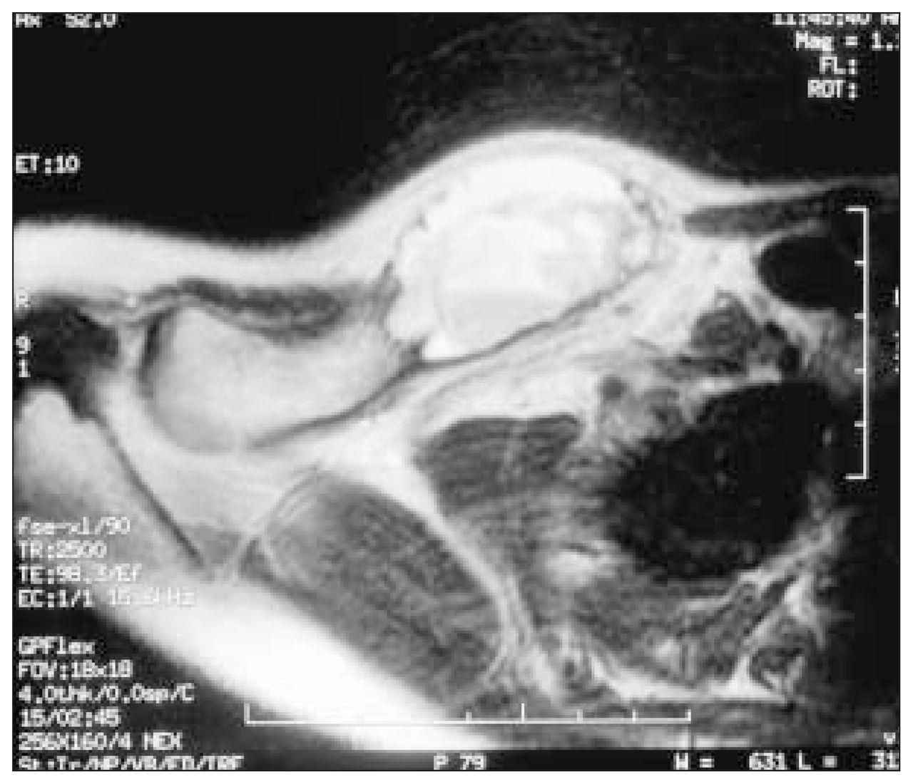
A plain radiograph of the clavicle is shown in Fig. 1 and a representative magnetic resonance image in Fig. 2. At operation, careful dissection revealed a cystic bluish mass with an extremely thin shell over overlying bone and periosteum (Fig. 3). The lesion was aspirated with a hypodermic syringe, and the lining was noted to collapse away from the surrounding bone. The lesion was entered and curettage done quickly to control bleeding. The periosteum was closed once hemostasis was ensured. The specimen was sent for pathological examination and culture.

A poorly defined lytic lesion involving the superior aspect of the middle third of the right clavicle (short arrow) with an associated supraclavicular soft-tissue mass (long arrow). There are no pulmonary or other bony abnormalities.

A fat-saturated, T2-weighted, axial magnetic resonance image shows a multicystic oval mass with a well-defined margin of thin cortical bone. The cyst content is layered, causing fluid–fluid levels. The high signal intensity layer is mainly plasma and the less intense gravity-dependent substance is due to red cells and cellular debris. The mass originates from the expanded middle third of the clavicle and the rest of the clavicle appears normal. Overlying soft tissue is displaced by the mass.

Intraoperative view of the aneurysmal bone cyst of the clavicle before removal.
The term aneurysmal bone cyst was popularized by Jaffe1 and Licht enstein2 in 1950 to describe a specific subperiosteal lesion. The lesions described were histologically characterized by large, cystically dilated vascular spaces, fibrosis, reactive bone, variable numbers of giant cells and scattered hemosiderin pigment. Radiologically they were characterized by a thin expandable shell of periosteal new bone overlying a superficially eroded outer metadiaphyseal cortex. These typically followed significant local trauma. Subsequently, this definition has been expanded to include intramedullary lesions with the same histologic characteristics.3 This terminology has caused considerable controversy, as many of these intramedullary lesions are the result of involutional changes in a number of different primary tumours.3 These primary tumours include simple bone cyst, giant cell tumour, osteoblastoma, chondroblastoma, angioma and telangiectatic osteosarcoma among others.4 Obviously considerable caution must be exercised in the diagnosis and management of the presumed aneurysmal bone cyst.
In the present case, the findings on radiography, computed tomography, magnetic resonance imaging and ultrasonography were all consistent with the presumed diagnosis of aneurysmal bone cyst. These demonstrated a cystic intramedullary lesion containing multiple septations. The overlying cortex had thinned and expanded.
Footnotes
Section editor: Robert S. Bell
Submissions to Surgical Images, musculoskeletal section, should be sent to Dr. Robert S. Bell, University Musculoskeletal Oncology Unit, Ste. 476, 600 University Ave., Toronto ON M5G 1X6; fax 416 586-8397
In this issue

Article tools


Related Articles
Cited By...
- No citing articles found.