


Presentation
A 43-year-old woman presented with a 5-day history of abdominal pain, fever and vomiting. There was no diarrhea, constipation or abdominal distension. She had undergone laparoscopic appendectomy 2 weeks before the onset of these symptoms. On examination, there was nonspecific abdominal tenderness. Bowel sounds were normal. The patient underwent plain abdominal radiography and contrast-enhanced CT of the abdomen. The representative images (Fig. 1, Fig. 2) are shown.
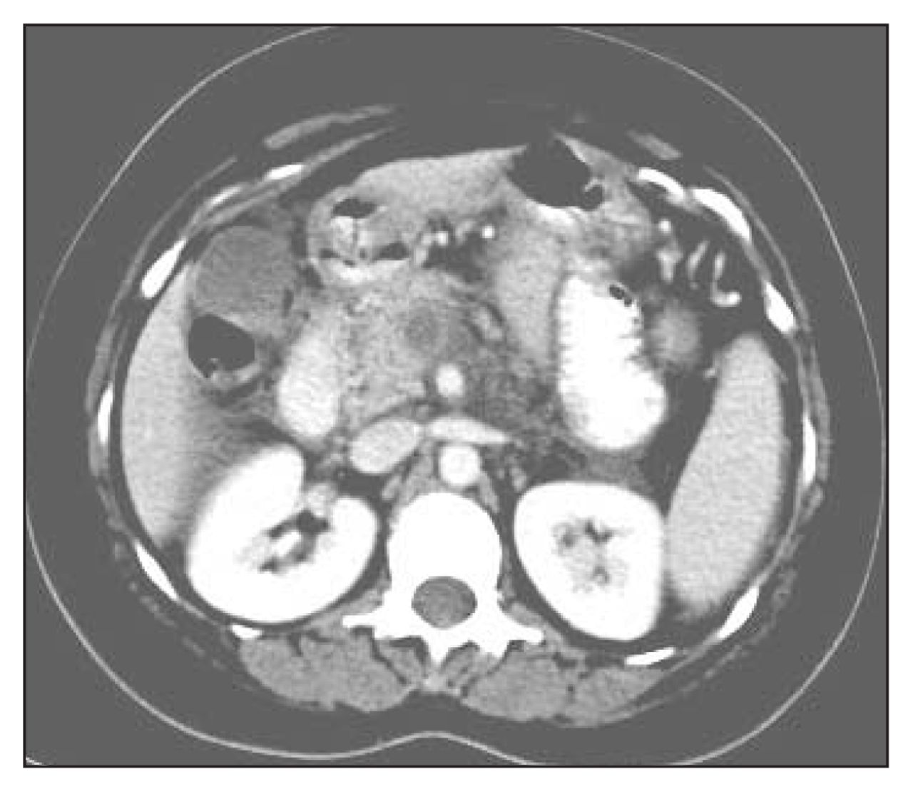
Axial contrast-enhanced CT image below the level of the pancreas.

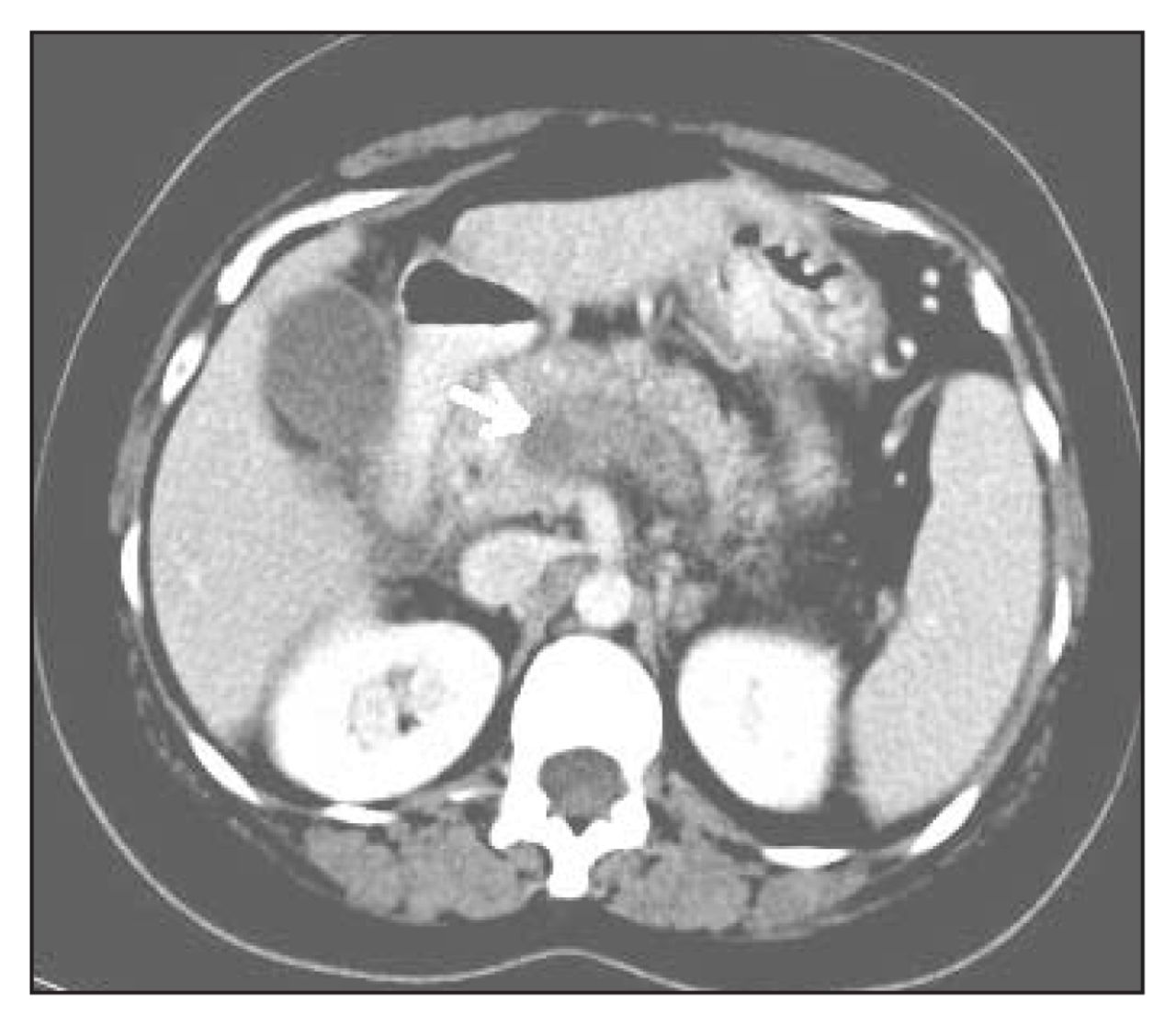
Axial contrast-enhanced CT image at the level of the pancreas.
What is your diagnosis?
Diagnosis
Superior mesenteric vein thrombosis
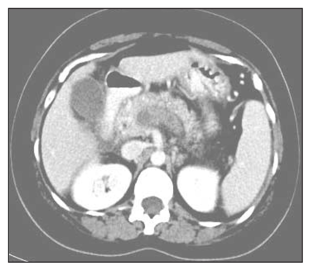
Axial contrast-enhanced CT images show a hypodense thrombus within the superior mesenteric vein (SMV) (Fig. 3, arrow) and the portal vein (Fig. 4, arrow). No abnormality was detected in the right iliac fossa. There was no evidence of air in, or adjacent to, the thrombus and no evidence of liver abscess. Thrombosis of the SMV and the portal vein following appendicitis and appendectomy was diagnosed. The patient received anticoagulant therapy, initially with heparin and subsequently warfarin. The findings on thrombotic work-up were negative.

A hypodense thrombus is seen within the superior mesenteric vein (arrow) on axial contrast-enhanced CT.
A hypodense thrombus is seen within the portal vein (arrow) on axial contrast-enhanced CT.
SMV thrombosis is a rare entity. It is a complication of appendicitis, and can follow appendectomy. Multiple factors may influence the development of SMV thrombosis after appendectomy, including hypercoagulation disorders, platelet abnormalities, rheologic changes and abnormalities of the splanchnic vessels.1 The complication is more frequent if the surgery is performed under difficult conditions (peritonitis) or when the patient presents with coagulopathy.2 Extension of the thrombus to the portal vein is associated with a higher risk of bowel ischemia.
The diagnosis of SMV thrombosis after appendectomy is usually not suspected clinically because the symptoms and the signs are nonspecific.3 CT has proven effective in the diagnosis and evaluation of SMV and portal vein thrombosis.4 With contrast-enhanced CT, SMV thrombosis is characterized by a decreased intraluminal density, occasionally combined with a ring enhancement of the vein, because of opacification of the vasa vasorum.1 These patients are treated with early anticoagulation. The mortality and morbidity associated with SMV thrombosis are high. Thrombolysis has recently been reported as an alternative therapy.5
Footnotes
Submissions to Radiology for the Surgeon, soft-tissue section, should be sent to the section editor: Dr. Lawrence A. Stein, Department of Radiology, Royal Victoria Hospital, 687 Pine Ave. W, Montréal QC H3A 1A1; lawrence.stein{at}muhc.mcgill.ca
Competing interests: None declared.
In this issue

Article tools


Related Articles
Cited By...
- No citing articles found.