

Bronchial Carcinoid
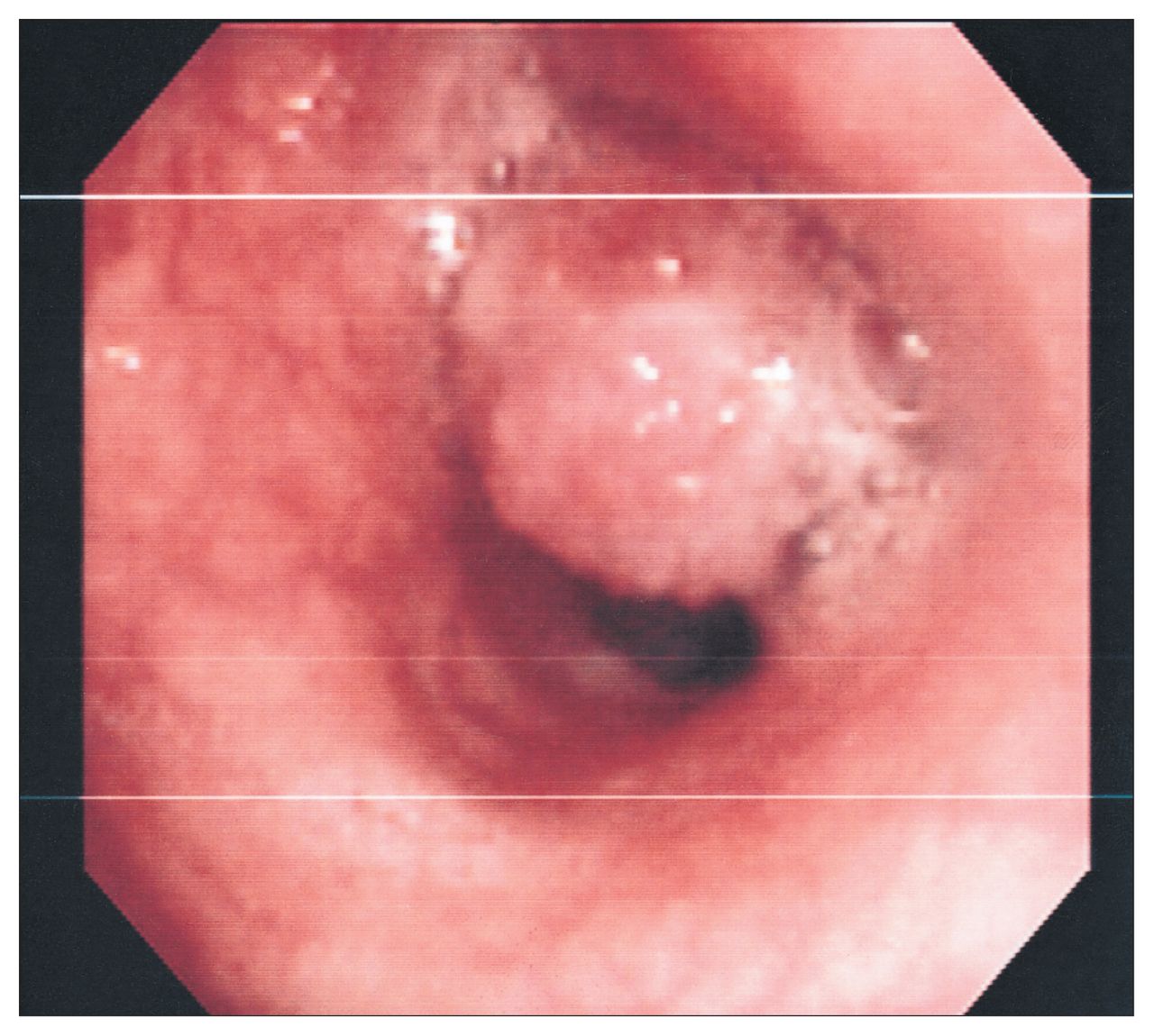
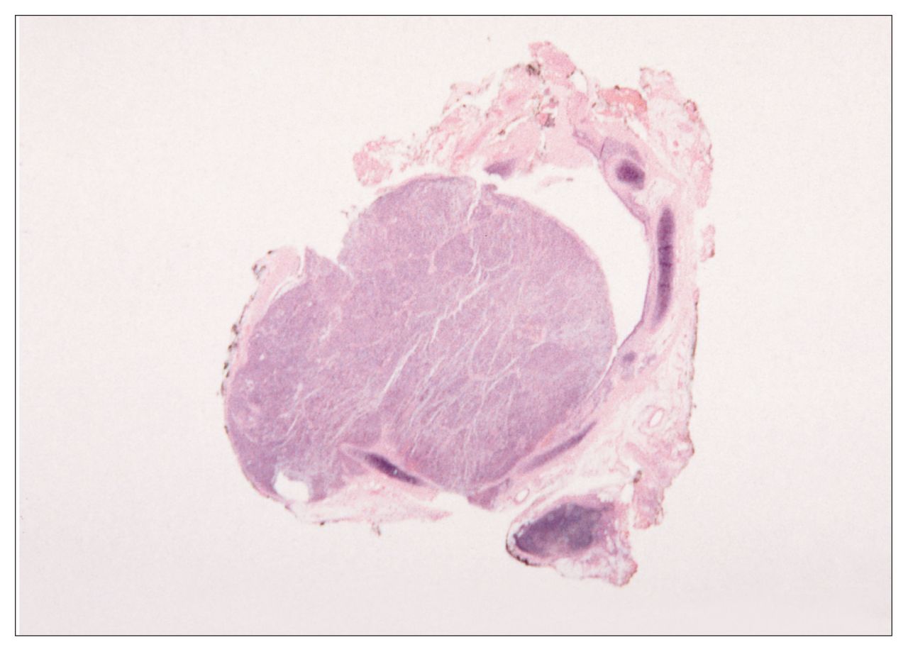
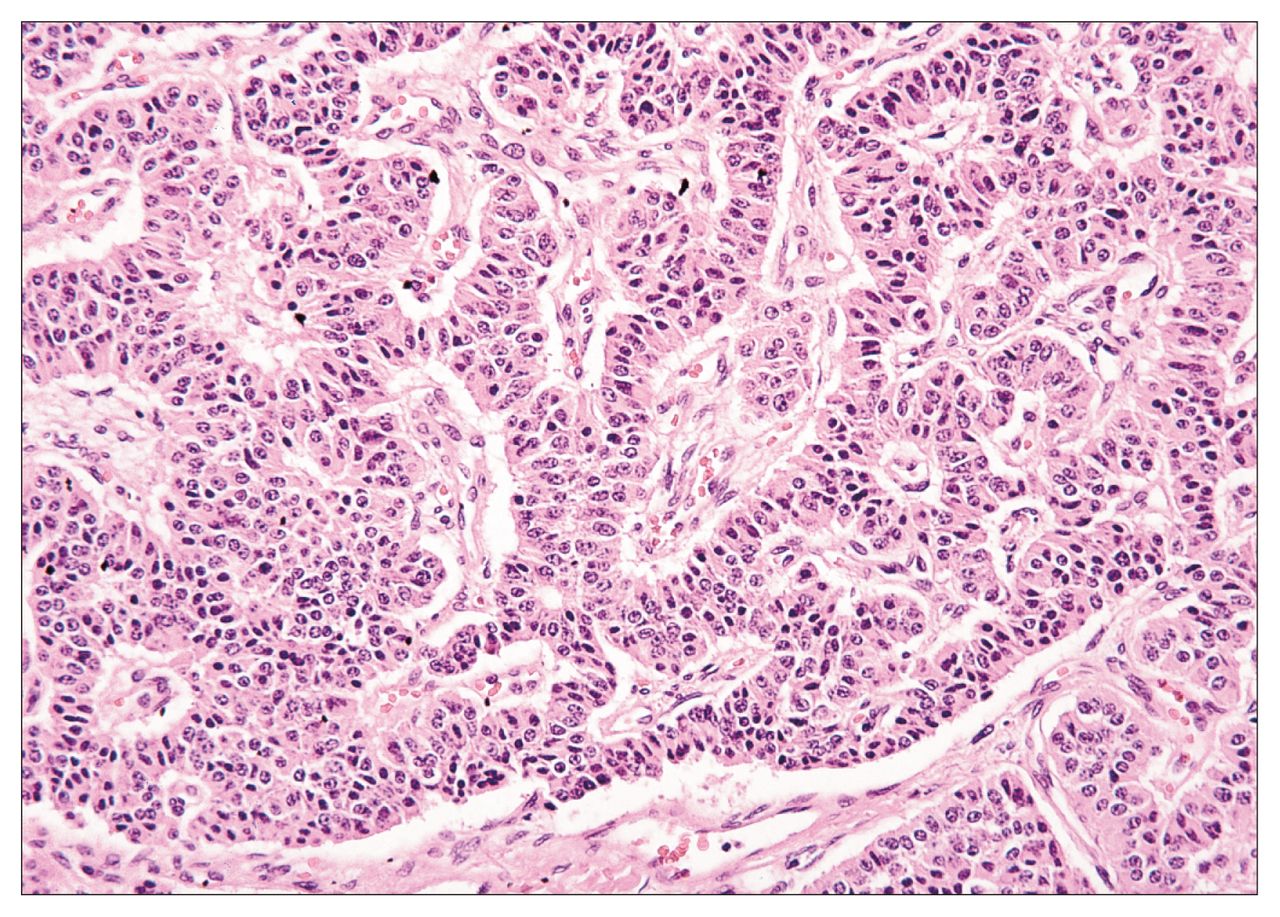
The chest computed tomography scan (Fig. 1) showed an endobronchial lesion (black arrow) completely obstructing the left lower lobe bronchus, causing collapse of the left lower lobe (white arrows). Bronchoscopy identified an obstructing polypoid lesion (Fig. 2) located at the bifurcation of the left upper and lower lobe bronchi. Fine-needle biopsy revealed typical carcinoid. The patient underwent an uncomplicated left thoracotomy and sleeve lobectomy of the left lower lobe. The gross specimen (Fig. 3) shows the 2.5 × 1.5 × 1.0-cm exophytic lesion nearly filling the bronchial lumen but only minimally infiltrating the bronchial wall. The semicircular rings of cartilage highlight the underlying architecture. Histologic examination (Fig. 4) revealed anastomosing trabeculae of uniform polygonal cells having round nuclei, granular chromatin, and inconspicuous cytoplasm with no significant cytologic atypia, few mitoses and no necrosis, characteristic of a typical bronchial carcinoid. The patient recovered fully and follow-up bronchoscopy showed a patent bronchial anastomosis.


