

Ultrasonography on a 25-year-old woman suffering from right upper quadrant pain and having mild jaundice revealed a complex cystic fullness of the subhepatic biliary system. Endoscopic retrograde cholangiopancreatography (Fig. 1) followed by magnetic resonance (MR) imaging (Figs. 2 and 3) confirmed the presence of a type I choledochal cyst with gallstones. Computer-generated 3-dimensional reconstruction using MR data created a unique virtual endoscopic view of the extrahepatic ductal (Figs. 4 and 5) and gallbladder (Fig. 6) anatomy.
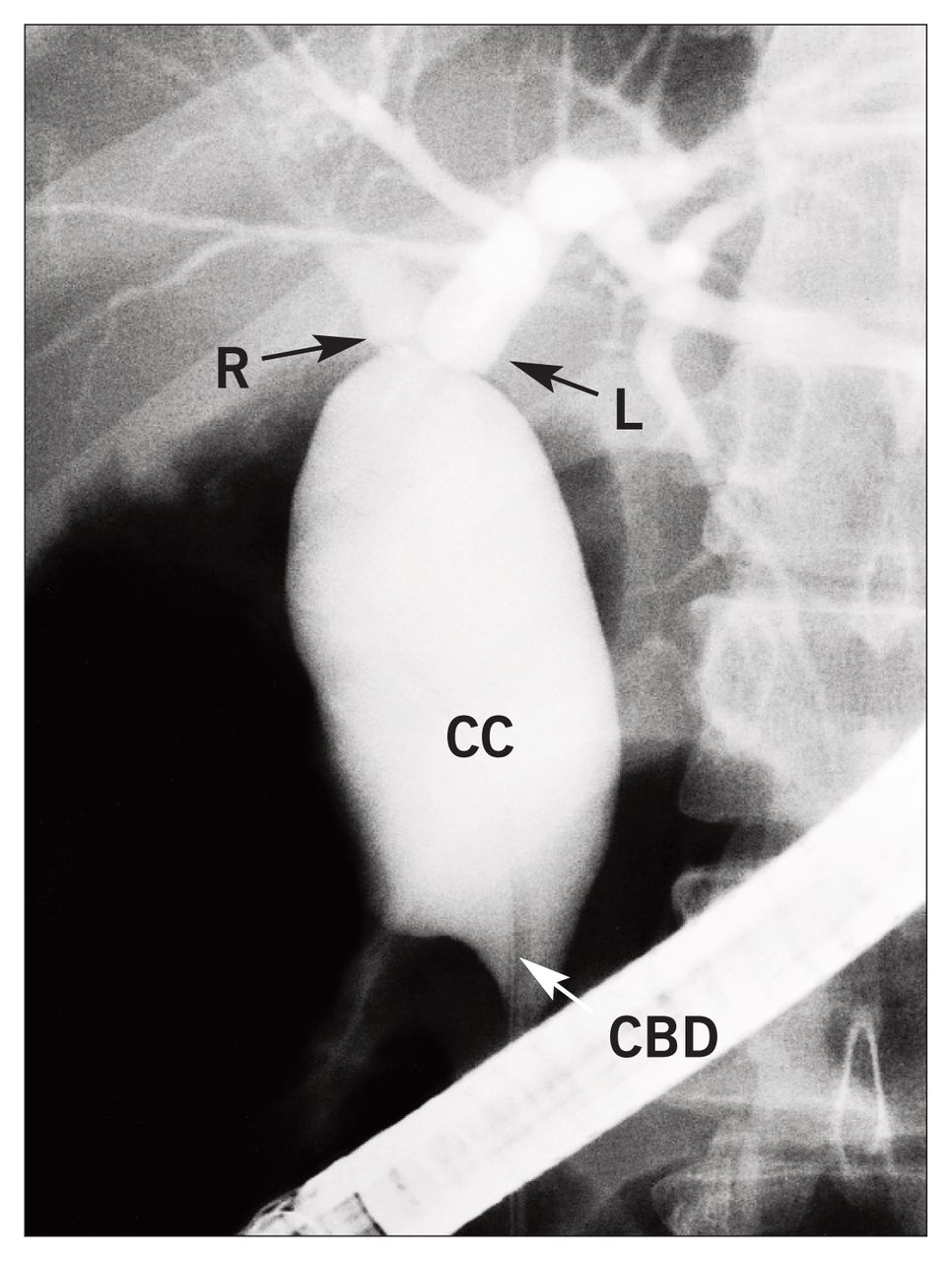
Endoscopic retrograde cholangiopancreatography of the choledochal cyst (CC). R = right hepatic duct, L = left hepatic duct, CBD = common bile duct.
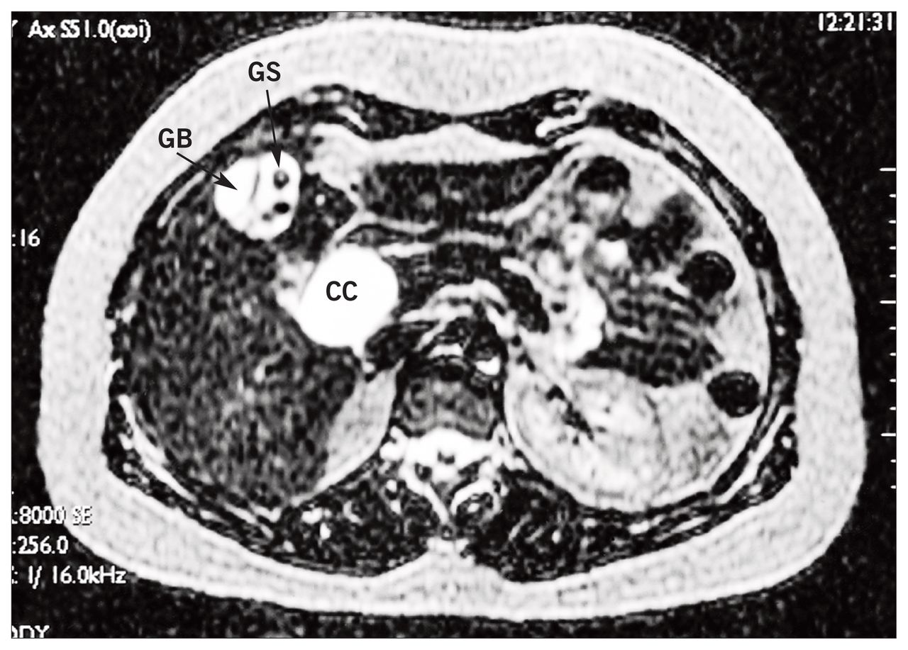
Transverse magnetic resonance image of the choledochal cyst (CC) and gallbladder (GB) with stones (GS). Heavily weighted axial T2 spin echo sequence.

Subtraction magnetic resonance image of the choledochal cyst (CC) and gallbladder (GB) with stones (GS) showing the hepatic ducts (R and L) and the distal common bile duct (CBD). Maximal intensity 3-dimensional projection (non contrast).

Computer-generated endoscopic view of the choledochal cyst (CC).

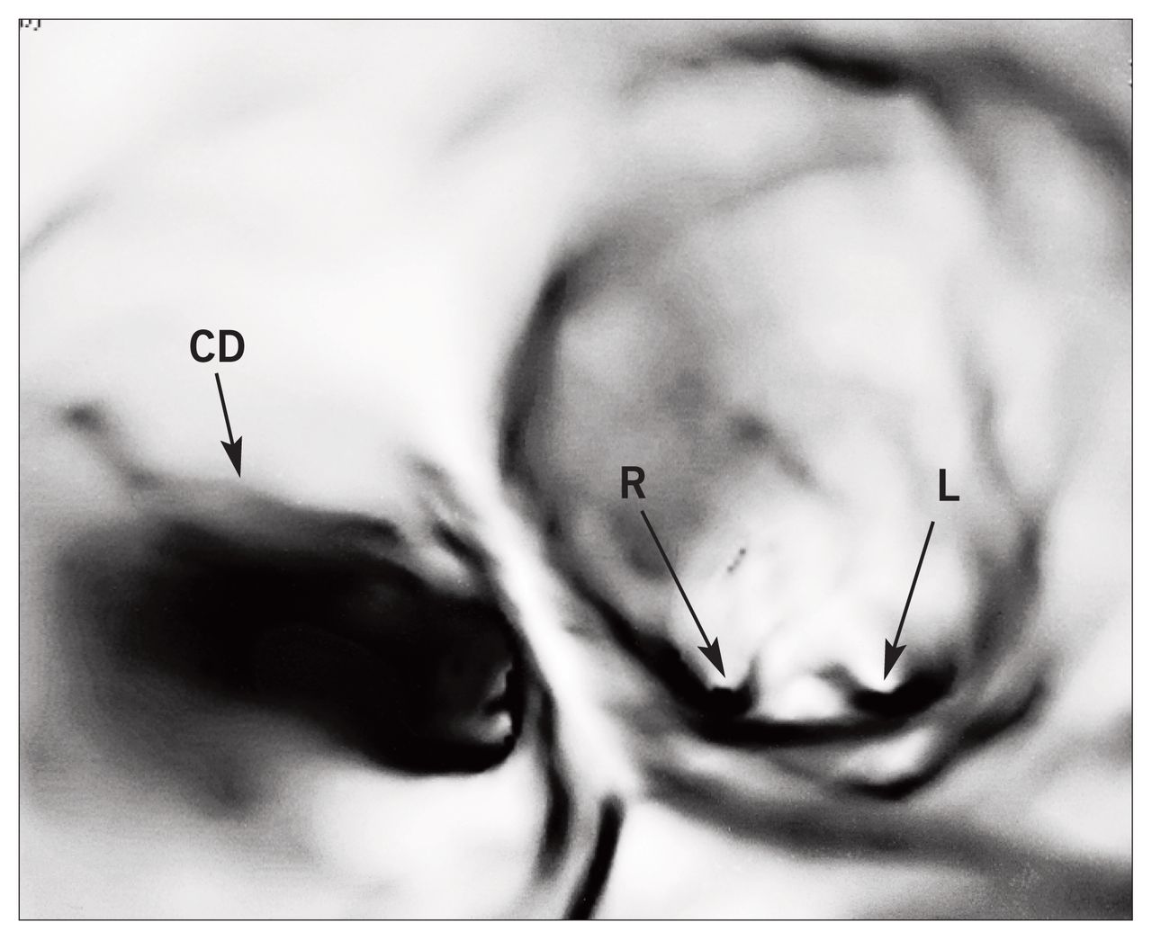
Computer-generated endoscopic view of the choledochal cyst and common hepatic duct at its bifurcation with the cystic duct (CD) and right (R) and left (L) hepatic ducts.
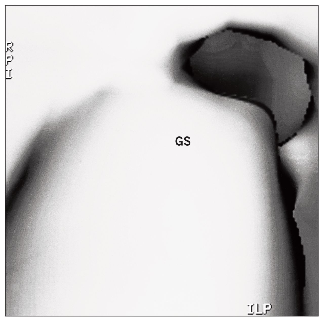
Computer-generated endoscopic cholecystogram showing a gallstone (GS).
Type I choledochal cysts are the most frequent congenital deformity of the common bile duct, which may not become symptomatic until adult life. Obstruction of the biliary tree because of stasis, stricture and stone formation favours septic complications and a higher incidence of malignant transformation (20 times that of the general population). This type of de formity requires resection and a Roux-en-Y choledocho(hepatico)jejunostomy.
Footnotes
Section Editors: David P. Girvan, MD, and Nis Schmidt, MD
Submissions to Surgical Images should be sent to Dr. David P. Girvan, Victoria Hospital Corporation, PO Box 5375, Station B, London ON N6A 5A5 or to Dr. Nis Schmidt, Department of Surgery, St. Paul’s Hospital, 1081 Burrard St., Vancouver BC V6Z 1Y6.