


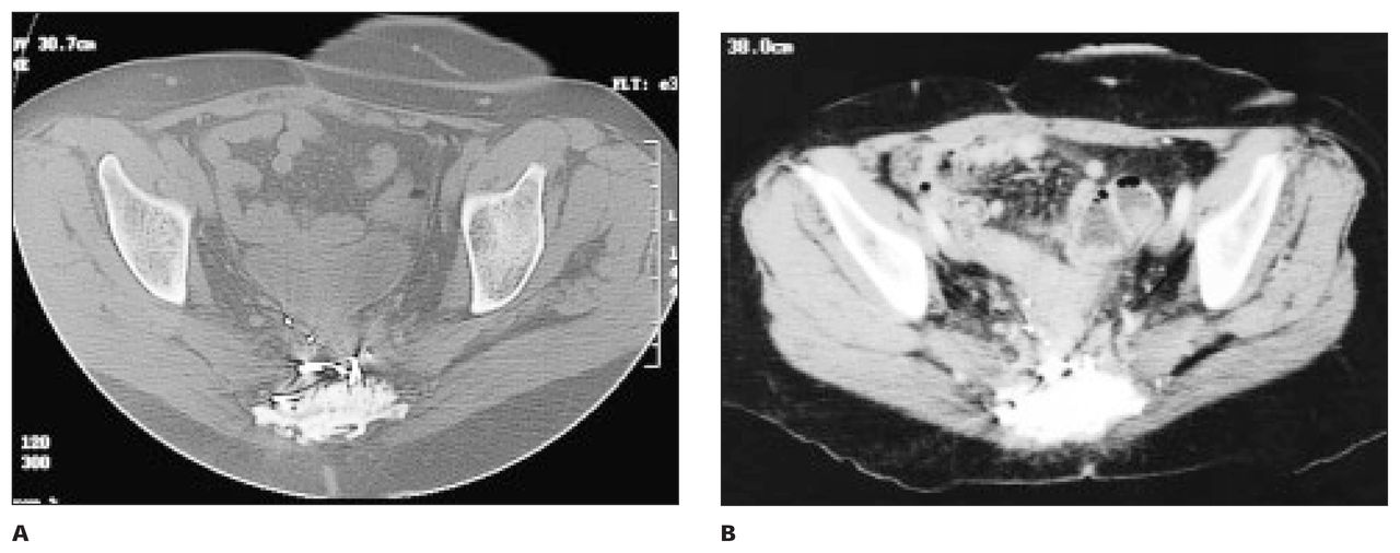
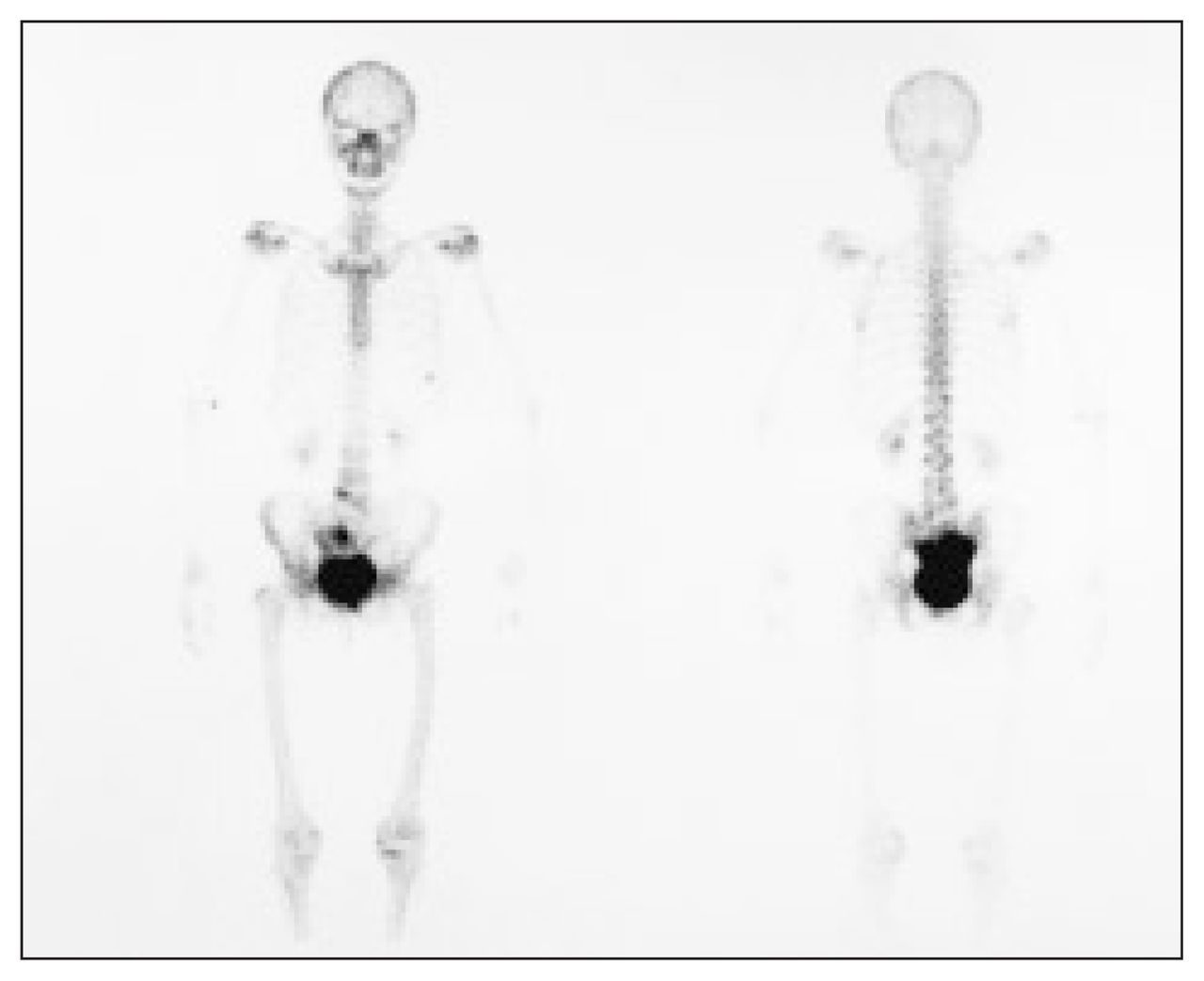
A 55-year-old man was referred to hospital because of sacral pain. He had a history of a rectal tumour that had been resected 10 years earlier and he had undergone adjuvant radiotherapy to the rectum. Over the last 6 months he had lost weight and suffered increasingly severe sacral pain. A plain radiograph of the pelvis showed increased density in the lower sacrum and coccyx and slight fragmentation inferiorly (Fig. 1). Surgical clips were noted lying anterior to the sacrum, in keeping with the history of rectal surgery. Axial computed tomography through the pelvis confirmed the markedly increased density of the sacrum with fragmentation inferiorly (Fig. 2). No associated soft-tissue mass was identified. More proximally, it was possible to identify the affected–normal sacral interface (Fig. 3). A colostomy was identified in the left iliac fossa (white arrow). An isotope bone scan demonstrated marked uptake of isotope within the sacrum, most marked posteriorly (Fig. 4).


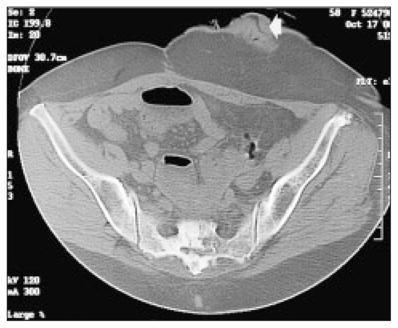

What is the diagnosis?
For the answer and discussion see page 346.
Footnotes
Section Editor: Peter L. Munk, MD
Inquiries about this section should be directed to Dr. Peter L. Munk, Professor, Department of Radiology, Vancouver General Hospital and Health Sciences Centre, 855 West 12th Ave., Vancouver BC V5Z 1M9.
In this issue

Article tools


Related Articles
Cited By...
- No citing articles found.