


Identification of secondary malignant change in a long-standing benign cartilage tumour of bone (enchondroma) can be difficult. One of the most important signs of malignant transformation is evidence of bone erosion peripherally around the benign calcified enchondroma.
A 72-year-old man complained of distal femoral and patellofemoral pain. X-ray films demonstrated lysis around the periphery of the enchondroma (Fig. 1). This finding mandated a biopsy, which showed evidence of both Paget’s disease (Fig. 2 left, large arrow) and benign cartilage tumour (Fig. 2 right, small arrow).


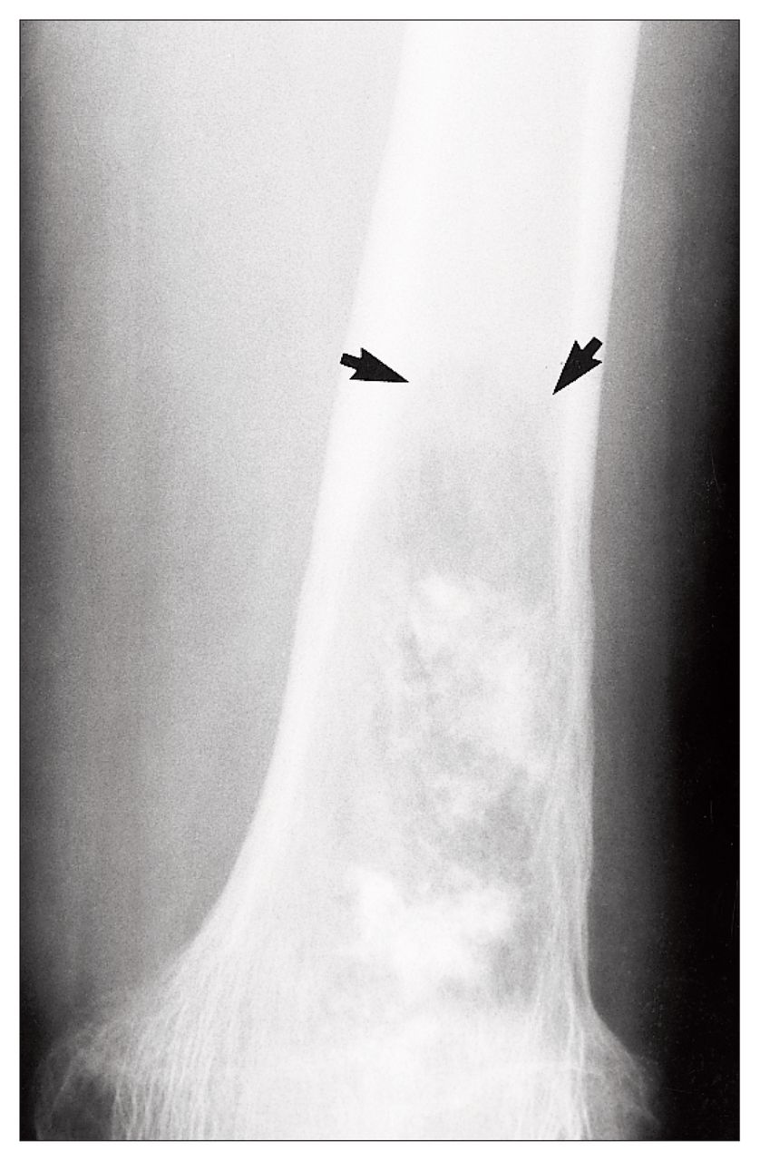
Follow-up x-ray films showed progression of the lytic change proximally in the femur (Fig. 3). This is the so-called “cutting cone” of progressive Paget’s disease. The patient had mild pain in the distal femur years after the initial presentation. The radiographic features of Paget’s disease typically move from the end of the bone toward the middle, and in this case migrated through a benign tumour.
Radiologic evidence of possible secondary malignant change around a benign cartilage tumour in this case resulted from Paget’s disease.
Footnotes
Section Editor: Robert S. Bell, MD
Submissions to Surgical Images, musculoskeletal section, should be sent to Dr. Robert S. Bell, University Musculoskeletal Oncology Unit, Suite 476, 600 University Ave., Toronto, ON M5G 1X5; fax 416 586-8397
In this issue

Article tools


Related Articles
Cited By...
- No citing articles found.