
Main menu

  • Home
  • Content
    • Current issue
    • Past issues
    • Sections
    • Collections
  • Podcasts
  • Author Info
    • Overview for authors
    • Publication fees
    • Forms
    • Editorial policies
    • Submit a manuscript
    • Open access
  • Careers
  • Alerts
    • Email alerts
    • RSS
  • About
    • General information
    • Staff
    • Editorial Board
    • Contact
  • CMAJ JOURNALS
    • CMAJ
    • CMAJ Open
    • JAMC
    • JPN

User menu

Search

  • Advanced search
CJS
  • CMAJ JOURNALS
    • CMAJ
    • CMAJ Open
    • JAMC
    • JPN
CJS

Advanced Search

  • Home
  • Content
    • Current issue
    • Past issues
    • Sections
    • Collections
  • Podcasts
  • Author Info
    • Overview for authors
    • Publication fees
    • Forms
    • Editorial policies
    • Submit a manuscript
    • Open access
  • Careers
  • Alerts
    • Email alerts
    • RSS
  • About
    • General information
    • Staff
    • Editorial Board
    • Contact
  • Subscribe to our alerts
  • RSS feeds
  • Follow CJS on Twitter
Radiology for the Surgeon

Soft-tissue case 31. Diagnosis

CAN J SURG February 01, 2000 43 (1) 22;
  • Article
  • Figures & Tables
  • Responses
  • Metrics
  • PDF
Loading

Emphysematous cystitis

The patient received antibiotics intravenously shortly after admission. Because of signs of peritonitis, laparotomy was done. The urinary bladder was found to be indurated and hemorrhagic, with crepitations around the wall. Cystoscopy showed a diffusely inflamed mucosa. Bladder lavage was done and a urinary catheter placed for drainage. Postoperatively the patient received antibiotics intravenously and was monitored in the intensive care unit. She suffered multiple organ failure and died 3 days after the operation. Culture of the urine grew Klebsiella pneumoniae.

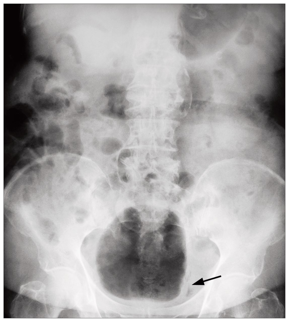
Emphysematous cystitis is a rare complication of urinary tract infection that usually occurs in diabetic patients. 1 Fermentation of glucose by bacteria causes gas production in the lumen and wall of the bladder, resulting in a rim of radiolucency around the bladder on plain films (Fig. 1).2 The common responsible organisms include Escherichia coli, Enterobacter and, rarely, Klebsiella and fungus.3 The condition is difficult to diagnose because presenting symptoms are diverse. Urinary symptoms and an unusual radiologic appearance may prompt earlier involvement of urologists. Early recognition, control of diabetes, aggressive intravenous antibiotic therapy and adequate bladder drainage are the mainstays of treatment. The outcome of this condition is variable, ranging from complete recovery to death, depending on the general condition of the patient and the severity of the illness at the time of presentation.3

FIG. 1
  • Download figure
  • Open in new tab
  • Download powerpoint
FIG. 1

References

  1. ↵
    1. Finby N,
    2. Begg CF
    . Diabetes mellitus and cystitis emphysematosa. N Y State J Med 1969;69(10):1315–8.
    OpenUrlPubMed
  2. ↵
    1. Bailey H
    . Cystitis emphysematosa: 19 cases with intraluminal and interstitial collections of gas. Am J Roentgenol 1961;86:850–62.
    OpenUrl
  3. ↵
    1. Quint HJ,
    2. Drach GW,
    3. Rappaport WD,
    4. Hoffman CJ
    . Emphysematous cystitis: a review of the spectrum of disease. J Urol 1992;147(1):134–7.
    OpenUrlPubMed
PreviousNext
Back to top

In this issue

Canadian Journal of Surgery: 43 (1)
CAN J SURG
Vol. 43, Issue 1
1 Feb 2000
  • Table of Contents
  • Table of Contents (PDF)
  • Index by author

Article tools

Respond to this article
Print
Download PDF
Article Alerts
To sign up for email alerts or to access your current email alerts, enter your email address below:
Email Article

Thank you for your interest in spreading the word on CJS.

NOTE: We only request your email address so that the person you are recommending the page to knows that you wanted them to see it, and that it is not junk mail. We do not capture any email address.

Enter multiple addresses on separate lines or separate them with commas.
Soft-tissue case 31. Diagnosis
(Your Name) has sent you a message from CJS
(Your Name) thought you would like to see the CJS web site.
CAPTCHA
This question is for testing whether or not you are a human visitor and to prevent automated spam submissions.
Citation Tools
Soft-tissue case 31. Diagnosis
CAN J SURG Feb 2000, 43 (1) 22;

Citation Manager Formats

  • BibTeX
  • Bookends
  • EasyBib
  • EndNote (tagged)
  • EndNote 8 (xml)
  • Medlars
  • Mendeley
  • Papers
  • RefWorks Tagged
  • Ref Manager
  • RIS
  • Zotero
‍ Request Permissions
Share
Soft-tissue case 31. Diagnosis
CAN J SURG Feb 2000, 43 (1) 22;
Digg logo Reddit logo Twitter logo Facebook logo Google logo Mendeley logo
  • Tweet Widget
  • Facebook Like

Related Articles

  • No related articles found.
  • Google Scholar

Cited By...

  • No citing articles found.
  • Google Scholar

Similar Articles

Content

  • Current issue
  • Past issues
  • Collections
  • Alerts
  • RSS

Authors & Reviewers

  • Overview for Authors
  • Publication Fees
  • Forms
  • Editorial Policies
  • Submit a manuscript

About

  • General Information
  • Staff
  • Editorial Board
  • Contact Us
  • Advertising
  • Reprints
  • Copyright and Permissions
  • Accessibility
  • CMA Civility Standards
CMAJ Group

Copyright 2023, CMA Impact Inc. or its licensors. All rights reserved. ISSN 2291-0026

All editorial matter in CJS represents the opinions of the authors and not necessarily those of the Canadian Medical Association or its subsidiaries.

To receive any of these resources in an accessible format, please contact us at CMAJ Group, 500-1410 Blair Towers Place, Ottawa ON, K1J 9B9; p: 1-888-855-2555; e: [email protected].

View CMA's Accessibility policy.

Powered by HighWire