


Right colonic intramural hematoma
The hematoma can be seen in Fig. 1. The patient was also investigated with small-bowel follow-through radiography, which showed that contrast passed by this lesion, which partially narrowed the lumen. The patient was treated nonoperatively and did not require laparotomy. He left hospital 3 days later tolerating a full diet. At follow-up he was well and had no complications (Fig. 2).

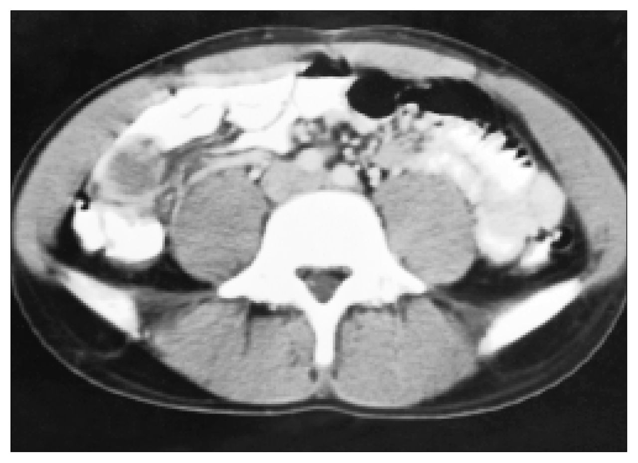
Computed tomography scan of the abdomen and pelvis with both oral and intravenous contrast media at the time of admission shows an intramural hematoma (arrow) of the cecum.
Follow-up CT scan 4 weeks later shows partial resolution of the hematoma.
Intramural hematomas have been described and reported to occur less frequently in the large bowel than the small bowel.1 Patients with these lesions are at risk for the development of stenosis in the long term, secondary to an organizing hematoma, and to bowel ischemia from vascular injury to the area.
A study by Rizzo and colleagues2 showed that CT is an accurate investigation for detecting bowel injury especially if oral contrast medium is used. In that study, CT findings that correlated with bowel or mesenteric injuries requiring surgery included the following: free peritoneal fluid, mesenteric infiltration, thick-walled bowel, free air and associated abdominal injuries. In patients treated non-operatively, CT scans showed bowel thickening but less frequently free peritoneal fluid, mesenteric infiltration and associated injuries.
CT of the abdomen and pelvis with both oral and intravenous contrast media is recommended for detecting injuries and hematomas of the bowel wall.
In this issue

Article tools


Related Articles
Cited By...
- No citing articles found.