


A 53-year-old woman presented to the Emergency Department on 2 occasions suffering from confusion and an altered level of consciousness. Hypoglycemia (2 mmol/L or less) was documented during these episodes. A 24-hour fast revealed symptomatic hypoglycemia (a blood glucose level of 2 mmol/L)and an elevated C-peptide level. At first, she was managed medically with frequent small meals and diazoxide. However, she continued to be symptomatic.
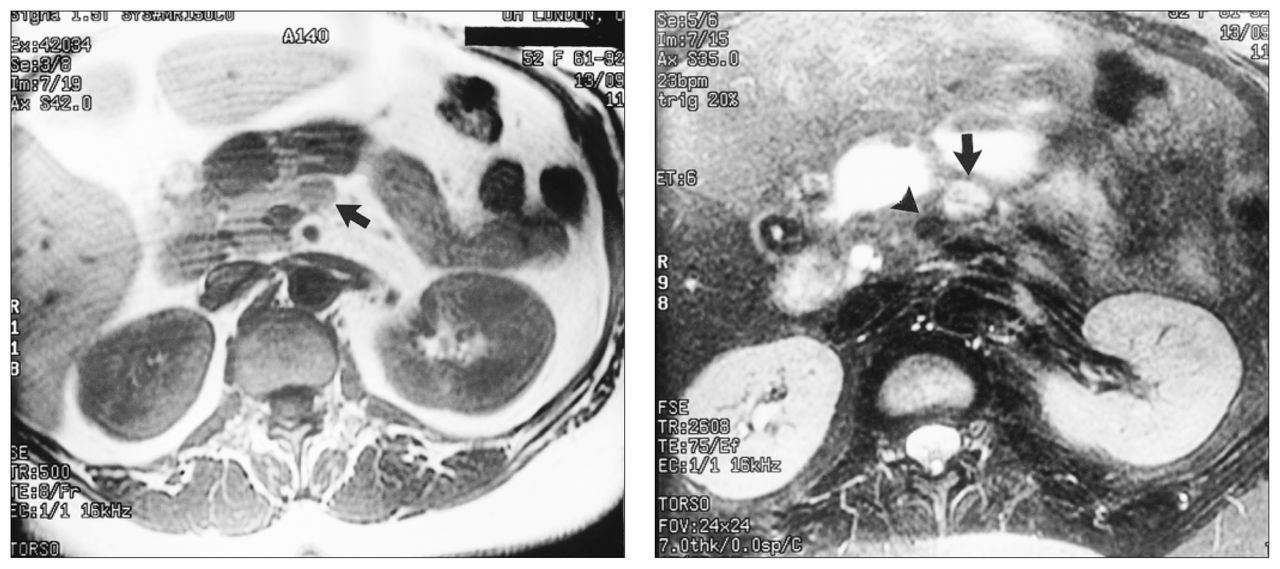
Two attempts to localize a suspected insulinoma by computed tomography and magnetic resonance imaging were unsuccessful. Finally, gadolinium-enhanced magnetic resonance imaging demonstrated a mass in the body of the pancreas measuring 1.5 cm in dimension (Fig. 1).

Left: T1-weighted image showing a hypointense mass in the pancreatic body (arrow). Right: the pancreatic mass becomes hyperintense on the T2-weighted fast spin-echo image (arrow). Note that the lesion is adjacent to, but not invading, the confluence of the splenic and portal veins (arrowhead). Pre-contrast and 30-second images from the gadolinium study showed rapid enhancement of the pancreatic mass (arrows).
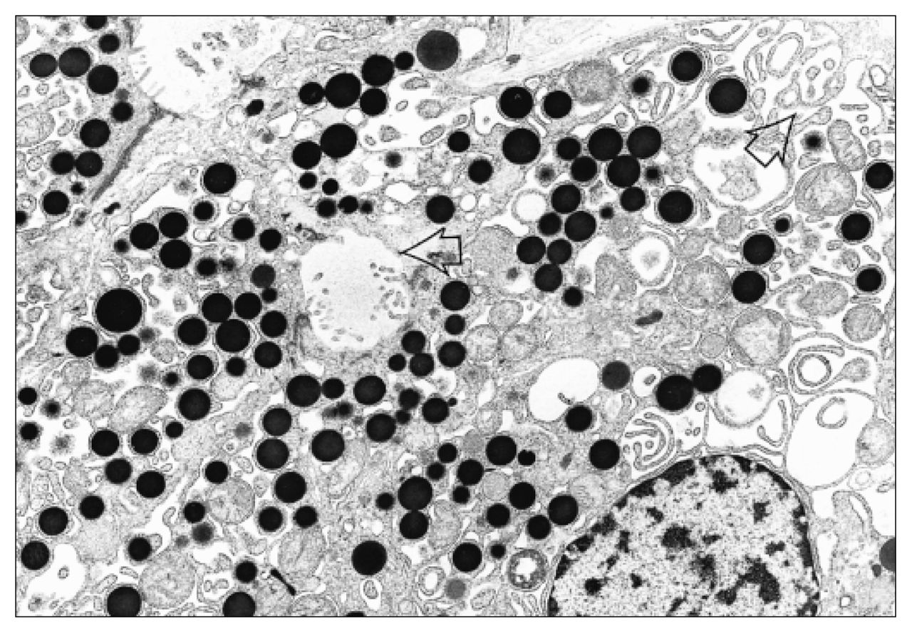
At laparotomy the pancreas was exposed; the mass (Fig. 2) was readily palpable. Although intraoperative sonography revealed that the mass was in close proximity to the splenic vein, the mass was enucleated without significant hemorrhage. Pathological examination supported the diagnosis of insulinoma. Electron microscopy showed abundant dense-core neurosecretory granules (Fig. 3) consistent with an islet cell tumour.
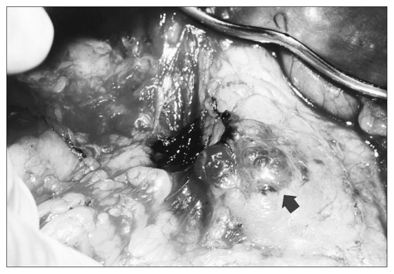
The intraoperative view clearly identifies the pancreatic lesion in the body of the pancreas (arrow).

Electron photomicrograph of the tumour shows numerous dense-core granules and microacini and microvilli (arrows) (original magnification × 7620).
Footnotes
Section editors: David P. Girvan, MD, and Nis Schmidt, MD
Submissions to Surgical Images, soft-tissue section, should be sent to Dr. David P. Girvan, Victoria Hospital Corporation, PO Box 5375, Station B, London ON N6A 5A5 or to Dr. Nis Schmidt, Department of Surgery, St. Paul’s Hospital, 1081 Burrard St., Vancouver BC V6Z 1Y6.
In this issue

Article tools


Related Articles
Cited By...
- No citing articles found.