


A 43-year-old woman was referred to the Department of Radiology for contrast enhanced magnetic resonance imaging (MRI) of her left shoulder and axillary region because of a palpable mass in her left axilla. She had initially noticed a lump 6 months previously while taking a shower. To her knowledge, the mass had not increased significantly in size during the intervening time. The mass was painless and caused no other symptoms.
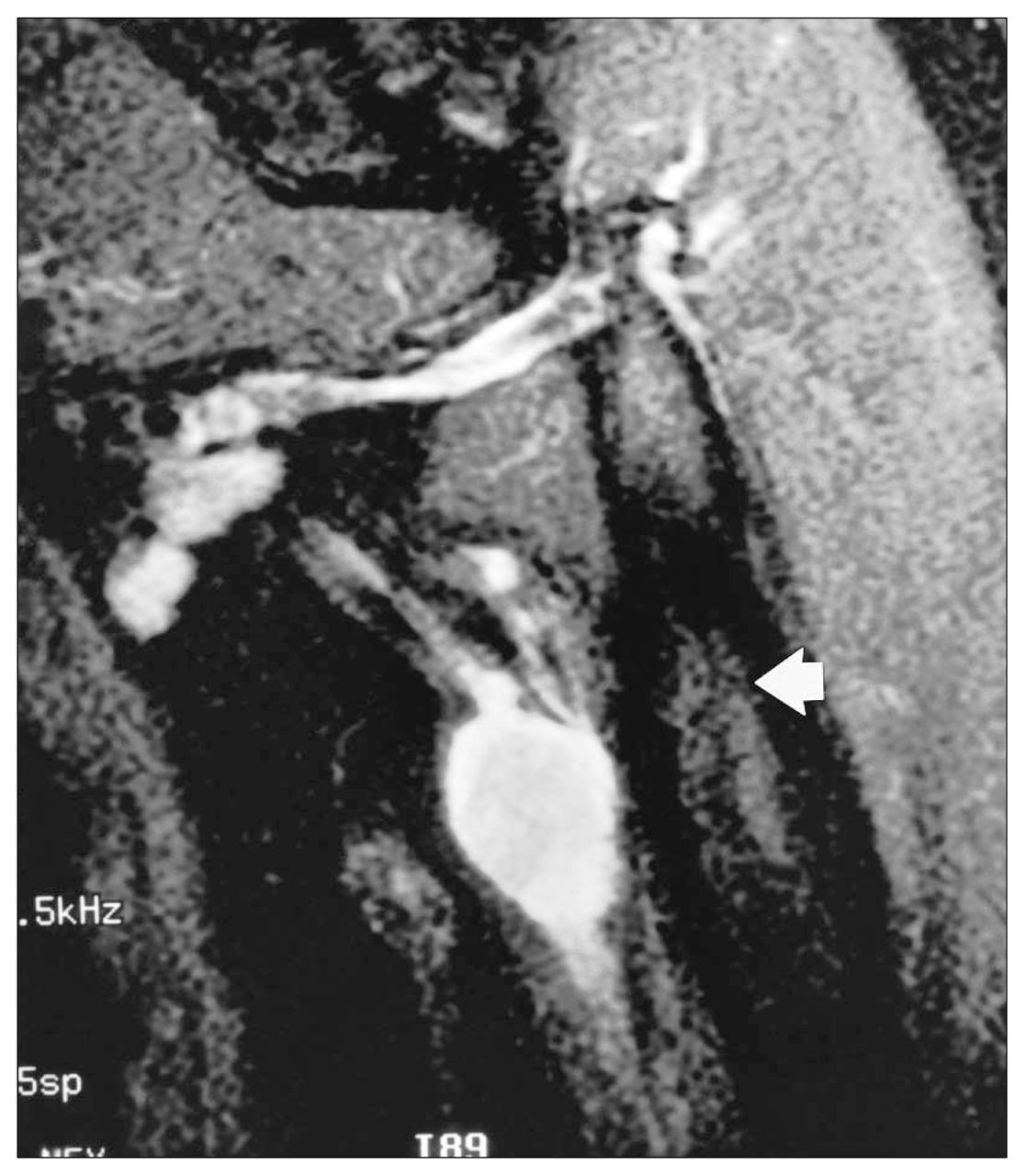
The mass was firm but mobile and appeared to extend in a cord-like fashion in direct relation to the neurovascular compartment of the axilla, extending inferiorly along brachial vessels. Axillary freckling was also noted. Several pigmented skin lesions were also seen scattered over the patient’s abdomen and back. Contrast enhanced MRI (Figs. 1 to 4) demonstrated a chain of enhancing masses of varying size, extending along the neurovascular bundle. Figs. 1 and 2 are contrast-enhanced T1-weighted axial images. Figs. 3 and 4 are fatsuppressed coronal T2-weighted images. Note that the humerus (arrow, Figs. 3 and 4) is of low signal intensity.


What is the most likely diagnosis?
For the answer and discussion see page 368.
Footnotes
Section Editor: Peter L. Munk, MD
Inquiries about this section should be directed to Dr. Peter L. Munk, Professor, Department of Radiology, Vancouver General Hospital, Vancouver Hospital and Health Sciences Centre, 855 West 12th Ave., Vancouver BC V5Z 1M9.
In this issue

Article tools


Related Articles
Cited By...
- No citing articles found.