


The patella is an uncommon site for primary bone tumours. The 2 commonest such tumours are giant cell tumour (GCT) and chrondroblastoma. We report a case of multicentric giant cell tumour involving the patella of a young adult with a previous GCT in the left fibula. Multicentric giant cell tumours are extremely rare and this is the first reported case, to our knowledge, of such a tumour involving the patella.
Case report
A 36-year-old woman had initially been seen in January 1987 because of a swelling on her left lower leg after a fall. The mass had increased slightly in size and was painful on palpation but not on walking. Radiography showed a lytic lesion within the proximal fibula, so she underwent resection of her left proximal fibula. She was seen in October 1996 complaining of persistent pain and a feeling of instability in her right knee. She dated the onset of the current symptoms to a minor fall and resultant injury to her right knee in April of that year, and she was treated with conservative management for what she was told was an undisplaced fracture of her patella. Review of the original radiograph obtained in April 1996 showed an undisplaced fracture at the superior pole of her right patella with possible underlying cystic changes (Fig. 1). A lateral view of the right knee obtained in January 1997 showed an enlarged patella and definite cystic changes within the bone (Fig. 2). Magnetic resonance imaging showed abnormal enlargement and sclerosis of the patella. The normal, bright T1-weighted marrow signal was replaced by a homogeneous low signal. On T2-weighted images, a linear high signal could be seen extending across the bone, consistent with a fracture (Fig. 3).

An oblique view of the patella showing an undisplaced fracture and possible underlying cystic changes.

A lateral view of the right knee showing an enlarged patella with multifocal lytic destruction of bone. The cortex is focally thinned along the endosteal surface.
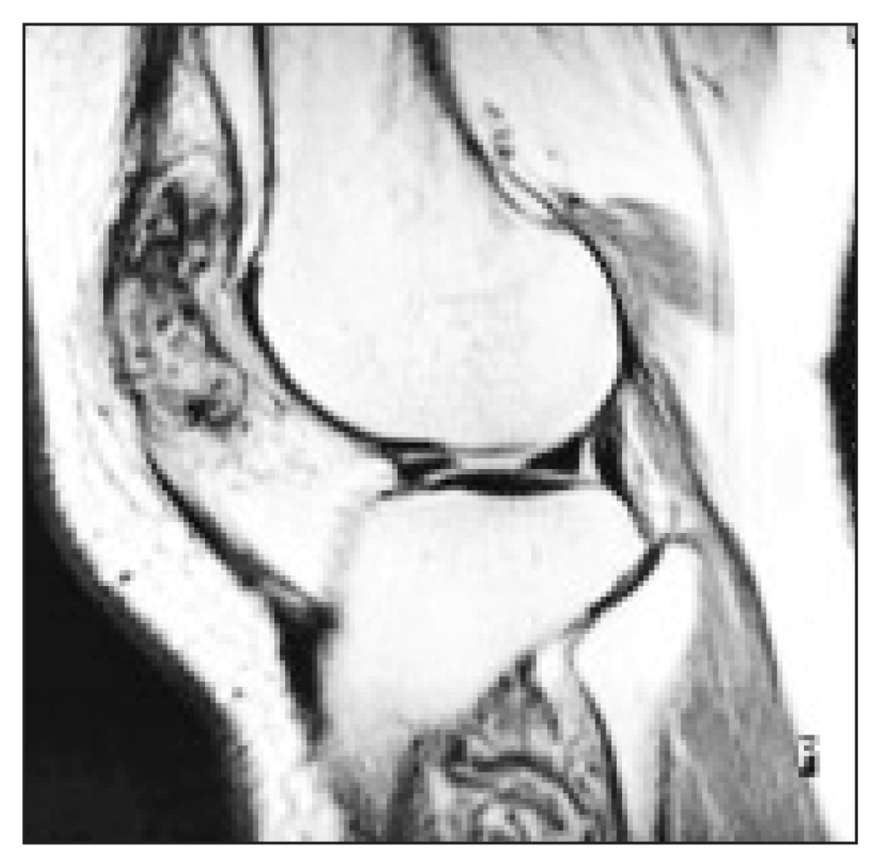
A magnetic resonance image with the lateral view of a T1-weighted sequence. The patella is enlarged and shows a uniform signal intensity. The cortex appears indistinct at several locations. No frank extension into adjacent soft tissue can be seen.
The radiologic appearance was considered unusual and the possibilities of a hematoma, secondary infection or a nonaggressive tumour of the patella such as a GCT, fibrous dysplasia or aneurysmal bone cyst were considered. The patient underwent arthroscopic examination of her right knee in March 1997. This demonstrated replacement of the articular cartilage by fibrous tissue on the inferior surface. The pain, however, persisted and repeat radiography, performed in April 1998, showed an enlarged patella with extensive lucent and sclerotic areas contained within it. A provisional diagnosis of fibrous dysplasia was made and a patellectomy was carried out 4 months later.
On examination of the operative specimen, the patella was enlarged, measuring 5.0 × 5.0 × 2.5 cm in dimension, and had a homogeneous, white appearance with fibrous adhesions. On cut section, the patella was replaced by brownish-tan tissue, which had a soft consistency with areas of hemorrhage. A specimen radiograph showed an expansile lytic lesion with irregular margins but no breakthrough of the cortex and no sclerotic reaction. No calcifications were seen within the lytic lesion (Fig. 4).

The surgical specimen showing an enlarged patella with replacement of the marrow cavity by a uniform softtissue mass. Endosteal scalloping and thinning is present but no obvious cortical breakthrough can be seen. One area of trabeculation is noted at the superior pole and may possibly relate to the site of the previous fracture.
Microscopically, the lesion showed the typical features of a GCT with a round to polygonal mononuclear cell proliferation containing numerous multinucleated giant cells. Reactive new bone formation was evident at the periphery of the tumour. The lesion did not invade the subchondral bone plate. Mitotic figures ranged from 2 to 7 per 10 high-power fields and some appeared asymmetrical. A fracture site could not be identified with certainty. Re-examination of the initial slides from l987 showed identical findings to the present lesion with a small focus of tumour at the superior resection margin. The patient made a smooth recovery and she was asymptomatic 15 months later.
There were no clinical features suggestive of hyperparathyroidism in this patient and calcium levels were reported as normal. The most recent bone scan showed no evidence of other skeletal lesions.
Discussion
Giant cell tumours make up 4% to 5% of primary bone neoplasms, with 40% to 50% reported to occur around the knee.1,2 Multicentric GCTs are extremely rare and account for less than 1% of all GCTs, with approximately 46 cases reported in the literature, the majority of these being single case reports.3–5 Multicentric GCTs present with clinical and pathological features similar to unifocal GCTs and most also occur around the knee.2 None of the 29 cases reported in the literature since 1972 and reviewed in detail by Cummins and associates3 were noted to involve the patella.
Pathologic fractures are not uncommon in GCTs of the patella and are reported to occur in 11% to 37% of patients. 2,5 They have also been reported to occur in greater frequency in patients with multicentric GCTs.3,4 The initial fracture noted in our patient’s first radiographic examination was probably not related to the trauma but rather to the presence of the underlying GCT.
Multicentric GCTs are reported to occur in a slightly younger population than typical, unifocal GCTs and are slightly more common in females.6 It is essential to exclude other possible causes of multiple lytic lesions such as multiple myeloma, hyperparathyroidism and metastatic carcinoma. Recurrent lesions may present up to 16 years after the first lesion, and in 15 of the 29 patients reviewed by Cummins and associates,3 a second lesion did not develop for more than 2 years after the initial presentation. In many patients there are multiple sites of tumour involvement. If a second lesion develops, as in this case, the patient is at increased risk of having further lesions in the future. Metastases to the lungs have been reported in 1% to 3% of cases of benign GCTs, with a common association of previous surgical intervention. 7 Patients with multicentric GCTs apparently do not have a higher incidence of pulmonary metastases.3 In a recent literature review of 39 patients with lung metastases of GCTs, only 4 patients died of their pulmonary disease.7
- Accepted July 3, 2000.
In this issue

Article tools


Related Articles
Cited By...
- No citing articles found.