


Presentation
A 35-year-old woman was referred to an orthopedic surgeon with a complaint of vague deep-seated pain in her right knee, which had been ongoing for a period of 3 months. There was no history of a specific inciting traumatic event.
Physical examination of her knee revealed no specific signs of internal derangement, and plain radiographs were unremarkable. The patient was referred for an MRI examination.
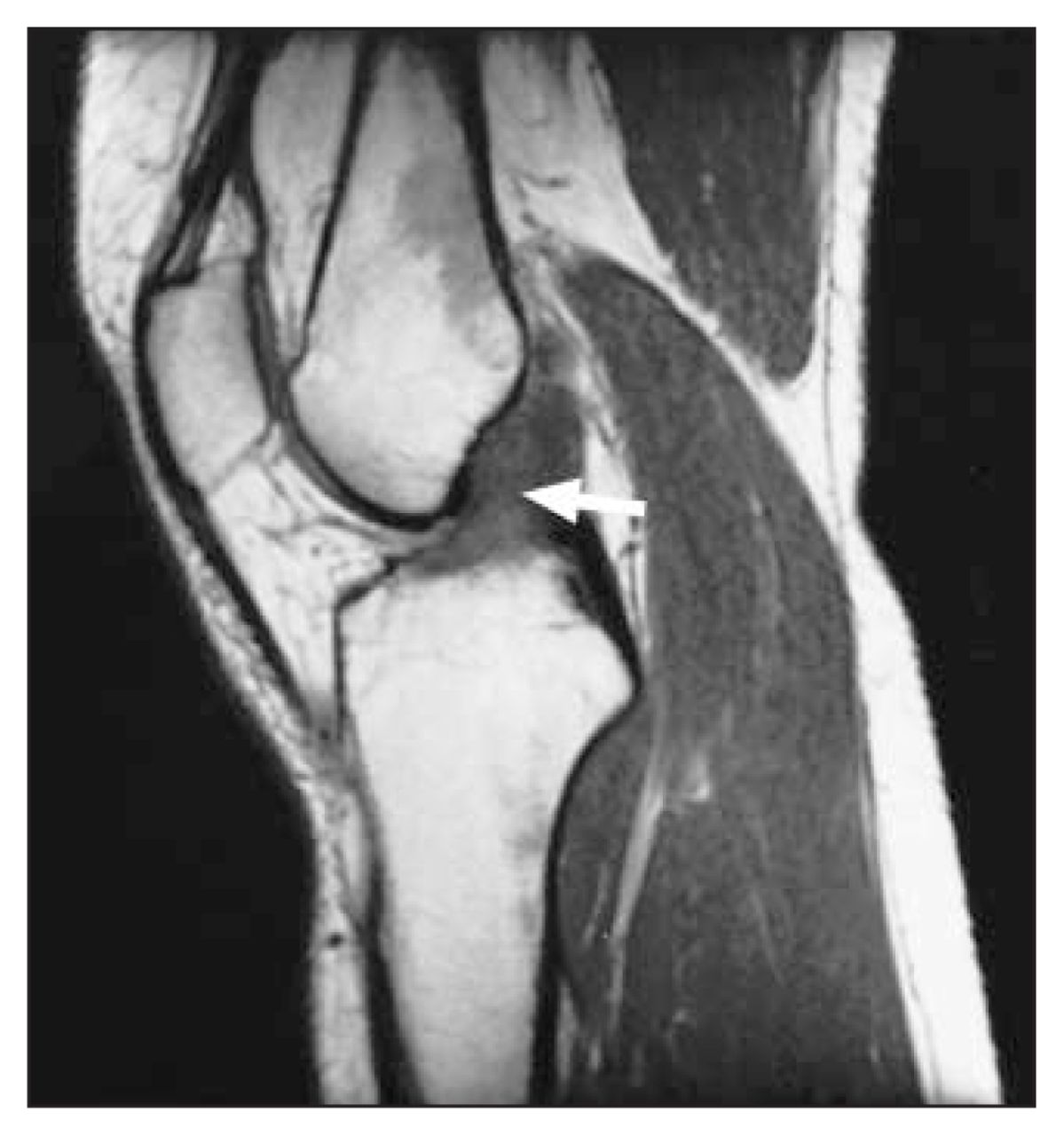
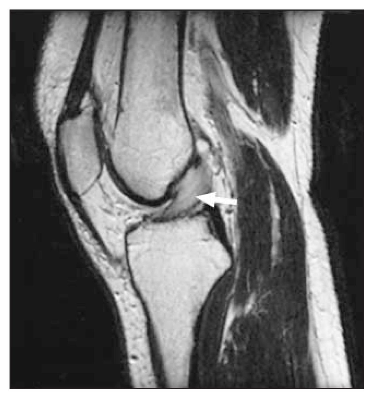
T1-weighted sagittal imaging demonstrated an isointense bulbous expansion of the anterior cruciate ligament (ACL) near its femoral attachment (Fig. 1, arrow). This bulbous expansion returned a high signal on T2-weighted imaging, which showed the lesion to be sited within the apical fibres of the ligament (Fig. 2). Axial fat-saturated T2 imaging confirmed the presence of the lesion (Fig. 3).


What is your diagnosis?
Diagnosis
Anterior cruciate ligament cyst
Cysts associated with the anterior cruciate ligament (ACL) are rare. Prevalence rates for cysts that are genuinely intra-ligamentous have been documented in two large MRI series as 0.25%1 and 0.44%.2 Similar rates have been noted for cysts related to the tibial and femoral insertion sites of the ligament.3,4
A cyst in the mid-portion of the ACL was first described by Caan in 1924,5 in the cadaver of an elderly man with no documented antemortem symptoms referable to the knee. The etiology of these lesions remains obscure, and a history of significant trauma is obtained in only a minority of cases.1 Theories include post-traumatic mucinous degeneration of connective tissue mediated by local release of hyaluronic acid, herniation of the synovium into a defect in surrounding tissue, and even displacement of synovial tissue during embryogenesis. 1,2 A strong male predominance exists. Symptoms comprise anteromedial knee pain aggravated by changing direction when running, on squatting or with extreme flexion and extension, and may resemble those of internal derangement.2
MRI, with its multiplanar capability, is the imaging modality of choice for diagnosis of these lesions, and demonstrates fusiform swelling of the ACL. The cysts return homogenously low signal intensity on T1- weighted images and high signal intensity on T2-weighted images, which are particularly good at contrasting the cysts against an intact ACL.1,2 The prevalence rate of associated internal derangement ranges from 22% to 50%.1,3
Most patients have good or excellent results after arthroscopic excision of ACL cysts; postsurgical recurrence has not been reported.3 Successful treatment with aspiration guided by computed tomography has also been described.6
Footnotes
Inquiries about this section should be directed to the section editor: Dr. Peter L. Munk, Professor, Department of Radiology, Vancouver General Hospital and Health Sciences Centre, 855 West 12th Ave., Vancouver BC V5Z 1M9; fax 604 875-4723; plmunk{at}interchange.ubc.ca
In this issue

Article tools


Related Articles
Cited By...
- No citing articles found.