


Dysplasia epiphysealis hemimelica (DEH), also known as Trevor’s disease, is a rare developmental disorder affecting the epiphyses; in young children, it usually involves the knee and ankle joints. DEH was first reported by Mouchet and Belot in 1926,1 who named it tarso megalia. In 1950, Trevor2 used the term tarso-epiphyseal aclasis. Fairbank3 in 1956 coined the term dysplasia epiphyseal hemimelica, the term that remains the common usage name for this unusual condition.
The etiology of DEH is unknown. Some authors have theorized that the defect is in the regulation of cartilage proliferation4 or that the disorder may be due to a localized disturbance of the pre–postaxial part of the apical cap of the limb bud in early fetal development.3 It does not appear to be genetically transmitted, 5–8 and no case of malignant transformation has been reported.6–10 The incidence of DEH has been estimated at 1 in 1 000 000.8 It is more common in males (3:1) and white Caucasians, and usually presents in childhood or early adolescence. The most common sites affected are around the knee, usually on the medial side of the tibial or femoral epiphysis, and the ankle, particularly the talus and the medial aspect of the fibular and tibial physis (Fig. 1). The natural history of DEH is that of continual growth of the lesion until skeletal maturity, at which time further enlargement ceases.

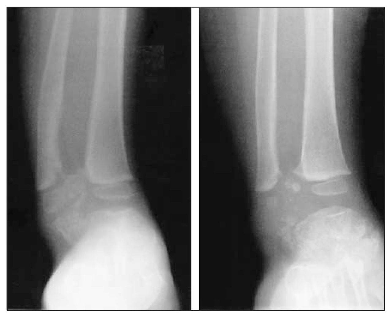
Anteroposterior (left radiograph) and lateral (right) views of patient A at 6 years of age, showing the typical medial involvement of the distal tibial epiphysis and medial talus. Note the misshapen contour of the medial part of the ankle.
The aim of this study was to examine the effects of DEH on ankle function and development and to recommend a treatment regime for this unusual but disabling condition.
Methods
Four patients with painful ankles, limited function and deformity secondary to DEH have been assessed at the Children’s Hospital of Eastern Ontario and the Winnipeg Children’s Hospital since 1985. Their surgical records, pathology reports, medical charts and radiographs before and after surgical excision were reviewed.
At surgery the protruding intra-articular cartilaginous mass was excised to restore joint congruity of the talus with the tibia and fibula, as well as to re-create the normal anatomy of the talus.
Results
All 4 children were boys, aged 1 to 4 years at presentation. Three were followed for an average of 3 years before surgical removal of the lesion. The other, with a lesion involving the fibula, underwent surgery shortly after diagnosis. Three required re-excision 5–7 years later. The longest follow-up was 11 years (patient C). Patient D is currently being followed subsequent to excision of a talar exostosis. The average follow-up from the first surgical treatment was 5.2 years; from diagnosis, 8.0 years. To date, none have required further surgery; patients A, B and C have exceeded skeletal maturity with no evidence of further growth of the lesions.
All had symptoms of pain in the ankle associated with running or long walks. Upon initial examination, all had restriction of dorsi-flexion and plantar flexion of the ankle of 10° or more compared with the contralateral side. Both ankle pain and loss of motion increased with time, correlating with increasing joint incongruity as the lesion grew. At the initial surgery, all children were noted to have “misshapen ankles” (Fig. 1). Surgical excision of the DEH lesion resulted in improved plantar and dorsiflexion in all children at each surgical intervention (Fig. 2) to a normal range of passive motion comparable to the contralateral normal ankle. All children returned to full unrestricted activity after surgery. The 3 boys who required a second procedure to restore joint congruity began experiencing a recurrence of pain and limitation of ankle motion 3–4 years after the initial surgery.

Left, an anteroposterior view illustrating exostosis from the fibula epiphysis (patient B, at age 1½ yr). Right, a resection 1 year postoperatively, with the exostosis removed and showing a more normal–appearing ankle joint.
All 3 children resumed their full physical education programs at school within 1 year of their second surgery and developed no later symptoms requiring further surgical intervention.
Discussion
Dysplasia epiphysealis hemimelica is a rare, unusual variant of osteochondroma primarily affecting the medial aspect of the epiphysis of the talus.
The 4 children in this review were all severely compromised by DEH affecting the ankle resulting in joint incongruity, limitation of ankle motion and pain after lengthy walking or running. Three of these children were followed for 3 years with attempted activity modifications and ankle supports, which were unsuccessful due to the relentless and progressive growth of lesions (Fig. 3).

Left, anteroposterior radiograph of the ankle of patient C at presentation (age 3½ yr). The abnormal articulation of the right fibula and talus is obvious, with the foot being pushed into varus by the fibular epiphyseal expansion. Right, an anteroposterior view of the ankle taken 4 years later at age 7½. Note the continued enlargement of the fibular epiphysis and the intra-articular exostosis from the talus.
Surgical removal of the lesions provided considerable subjective and functional improvement in ankle function. Based on this review of 4 children, early removal of DEH lesions is recommended even though complete extirpation of the lesion is usually impossible without excising a segment of the talus or epiphysis large enough to result in permanent compromise of the ankle joint. The objective of the surgery should be to remove the joint incongruity caused by DEH to facilitate smooth function of the articulation, with the realization that the procedure may have to be repeated once or twice before skeletal maturity, when the continued growth of DEH appears to cease. Although 3 of these cases predated the availability of magnetic resonance imaging, imaging the ankle annually to ascertain the extent of cartilage growth and asymmetry secondary to DEH is recommended, so that the developing incongruity can be surgical corrected before secondary adaptive changes occur in the joint.
Conclusions
Dysplasia hemimelica epiphysealis, although rare, causes considerable disability in ankle function due to joint incongruity caused by the lesion’s relentless growth during childhood. Early surgical removal to improve joint incongruity is recommended, with subsequent surgery to ensure continued congruity as required. Complete removal is usually not achievable initially. Children should be followed yearly, both clinically and with MRI, until the lesion stops growing at skeletal maturity.
Footnotes
Competing interests: None declared.
- Accepted August 26, 2004.
In this issue

Article tools


Related Articles
Cited By...
- No citing articles found.