


A 54-year-old woman presented to the emergency department with acute right lower quadrant abdominal pain of a few hours duration. Past medical history revealed recurrent lower abdominal pains, investigated 5 years previously by endoscopy and abdominal imaging, which were reportedly normal. On physical exam, there was an ill-defined mass in the right lower quadrant without any residual pain or other abnormal findings. Complete blood count and standard biochemical studies were normal.
Computed tomography of the abdomen showed an 11 cm × 3.8 cm fluidfilled appendix with intramural calcification (Fig. 1). There was a suspicious 2.3 cm lymph node in the right mesocolon. There was also a benign appearing cyst of the right ovary. On the basis of these findings, a diagnosis of mucocele of the appendix due to a possibly malignant lesion was made and the patient taken electively to the operating room a few days later for right hemicolectomy and right oophorectomy. During laparotomy, a dilated appendix was identified; however, it was homogenous and smooth, and it was not adherent to surrounding viscera. There was a small retracted lesion in the terminal ileum, with a desmoplastic reaction within the mesocolic and retroperitoneal tissues around the cecum. The patient underwent the procedure as planned, and postoperative course was uneventful.
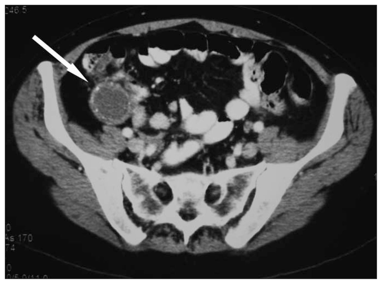
Computed tomography showing dilated appendix with intramural calcifications (arrow).
Histological examination showed a benign mucocele of the appendix secondary to mucosal hyperplasia and a 1-cm malignant carcinoid tumour of the terminal ileum, with had spread to 5 of 9 lymph nodes. Serial measurements of 5-hydroxyindoleacetic acid levels, computed tomography of the abdomen, positron emission tomography and octreotide scintigraphy remain negative at a follow-up of 20 months.
Discussion
According to the World Health Organization, 1 mucocele of the appendix may be divided into simple mucocele, resulting from hyperplastic epithelium or obstruction of the lumen; benign cystadenoma, secondary to mucus secretion by a benign adenoma; and cystadenocarcinoma, secondary to mucus-secreting adenocarcinoma of the appendix. In addition, it may be caused by a lesion known as mucinous tumour of indeterminate malignant potential.
In planning treatment, it is important to assess the likelihood of malignancy. Stocchi and colleagues2 reviewed 135 operated patients with mucocele of the appendix and found that the presence of symptoms, a palpable abdominal mass, a preoperative diagnosis (as opposed to an incidental intraoperative finding) and the presence of extravasation of mucus or pseudomyxoma peritonei correlated with the likelihood of malignancy. Dean and others3 also suggested a relation between the presence of abdominal pain and the likelihood of malignancy; however, in Stocchi’s review, cystadenoma was significantly larger than simple mucocele, and no lesion smaller than 2 cm was neoplastic. There was no correlation between size and the likelihood of malignancy per se. They recommend right hemicolectomy when malignancy is suspected. Pickhardt and colleagues4 also found that simple mucocele was rarely larger than 2 cm. In their analysis, intramural calcifications on abdominal imaging were highly suggestive of a neoplastic process, although not necessarily a malignant one.
Stocchi and colleagues found close to a 30% incidence of synchronous abdominal tumours, which is consistent with other authors.2 Most of these were colorectal malignancies. In our case, there was a synchronous carcinoid tumour of the terminal ileum, which had not been diagnosed on preoperative imaging and was found incidentally at laparotomy. We have found only one other such association in the literature.3 Abdominal exploration was otherwise negative. Our patient underwent upper and lower gastrointestinal endoscopy, and both were negative. Colonoscopy was not carried out preoperatively, to avoid blowing out the appendix and causing pseudomyxoma peritonei, although we are not aware of any reports of this type of complication.
In summary, it is important to recognize the entire spectrum of appendiceal disease that may present as a mucocele. Treatment is based on the likelihood of malignancy. It is also important to rule out synchronous abdominal tumours in this group of patients.
Footnotes
Competing interests: None declared.
- Accepted August 9, 2005.
In this issue

Article tools


Related Articles
Cited By...
- No citing articles found.