


Bone leiomyoma is a rare neoplasm, and only about 2 dozen cases have been reported in the literature.1–3 In this brief report we describe a case of leiomyoma arising from the iliac bone in an adult woman.
Case report
A 28-year-old woman with unremarkable medical history presented with a firm, painless mass on her right hip that had been present for 3 months. Physical examination and diagnostic imaging studies, including plain films and CT scans of her pelvis, confirmed the presence of a multilocular, osteolytic lesion on the upper part of her right iliac bone, measuring 7 cm in greatest dimension. The tumour was well circumscribed and incompletely surrounded by a thin rim of sclerotic bone (Fig. 1). Complete clinical investigation revealed no malignant tumour arising at other anatomic sites. Routine blood work and biochemical tests, including parathyroid hormone levels, showed no abnormalities. The lesion was subsequently removed by partial resection of the right iliac bone. Grossly, the resected bone fragment measured 13 × 11 × 3.5 cm and was surrounded by soft tissue. Serial sectioning of the specimen displayed an osteolytic firm tumour measuring 7 cm in greatest dimension. The tumour had destroyed overlying periosteum and expanded into the adjacent soft tissues with a pushing-type border.

Plain film of the patient’s right iliac bone reveals a multilocular osteolytic lesion without invasion of adjacent bone and partially surrounded by a rim of sclerotic bone (arrow).
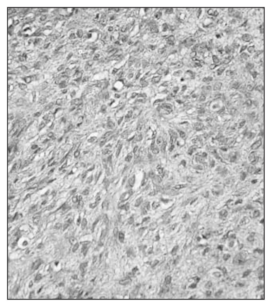
Histologically, the tumour was well-circumscribed, nonencapsulated and showed no invasion into the surrounding bone and connective tissues. It consisted of large bundles of spindle-shaped cells with eosinophilic, granular cytoplasm and uniformly elongated, normochromatic nuclei with small nucleoli. Neither mitotic figures nor tumour necrosis were identified (Fig. 2). Focal myxomatous change was noted elsewhere. The histologic pattern of the tumour was in keeping with a leiomyoma, and this was further supported by histochemical, immunohistochemical and ultrastructural studies of the tumour tissue. The patient was alive and well and showed no evidence of postoperative tumour recurrence or metastasis after 7 years.

Histologic view of the iliac leiomyoma shows solid sheets of spindle-shaped cells without nuclear atypia and mitotic figures (hematoxylin–eosin stain, original magnification 250 ×).
Bone leiomyoma is much less common than bone leiomyosarcoma, which accounts for about 0.7% of all primary bone cancers.4 Of the 24 cases of bone leiomyoma reported by 2004, 16 arose from gnathic bones and 8 from the remaining skeleton.1–3 Those tumours showed no bone invasion, were most commonly surrounded by a rim of sclerotic bone and displayed no cellular atypia or mitotic figures.1–3 Some had a prominent vascular component.1 The presence of a rim of sclerotic bone was not a reliable indicator of benignity, as it was seen in some cases of low-grade bone leiomyosarcoma.5 Thus, an absence of cellular atypia and mitoses in a smooth-muscle bone tumour, a noninvasive margin without bone destruction and the presence of a sclerotic bone rim around the tumour constitute important diagnostic criteria for a bone leiomyoma.1–3
Footnotes
Competing interests: None declared.
- Accepted April 10, 2006.
In this issue

Article tools


Related Articles
Cited By...
- No citing articles found.