


Chylothorax is a rare but recognized complication of esophageal surgery. The close anatomic relation between the esophagus and thoracic duct explains the risk of chylothorax after esophagectomy. We report a rare case of a thoracic lymphocele associated with a chylothorax after radical esophagogastrectomy. This case is unique in that, after a period of conservative management, a large lymphocele developed in the posterior mediastinum and compromised respiratory function. This led to surgical excision of the lymphocele and ligation of the thoracic duct.
Case report
A 56-year-old man was referred with a 3-month history of dysphagia to solids. Barium swallow examination demonstrated a 3-cm stricture of the distal esophagus, and esophagogastroduo-denoscopy (EGD) revealed a stenosis at the 25-cm level. Biopsy specimens of the lymphocele showed well-differentiated esophageal adenocarcinoma. Computed tomography (CT) of the abdomen and chest showed no evidence of metastases.
A 2-stage Ivor-Lewis radical esophagogastrectomy and 2-field lymphadenectomy were performed with reconstruction using a stapled esophagogastric anastomosis through the posterior mediastinum. A feeding jejunostomy and 2 right-sided thoracostomy tubes were placed.
Postoperatively on day 3, a chylothorax developed after the start of jejunal feeding, with chyle draining from the thoracostomy tubes. Pneumonia developed on day 14, and the patient required ventilation. Over this period, drainage from the thoracostomy tubes continued (average 2 L/d). Radiologic study with meglumine diatrizoate showed no evidence of anastomotic leakage. A left-sided pleural effusion, seen on chest ultrasonogaphy, was managed with a thoracostomy tube.
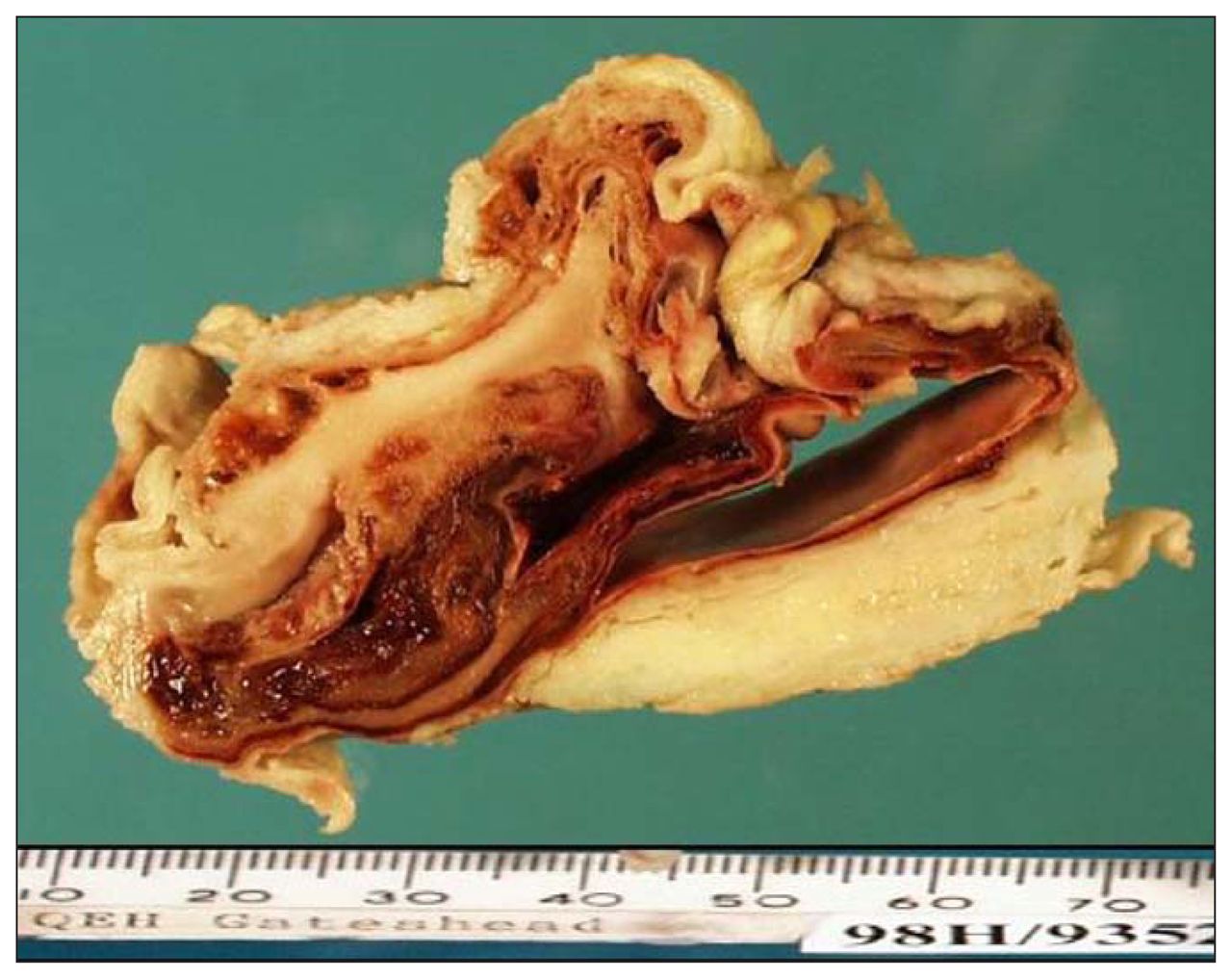
On day 37, the patient suffered respiratory failure with septicemia caused by methicillin-resistant Staphylococcus aureus, so vancomycin therapy was begun. A chest radiograph demonstrated a large opacification at the base of the right lung. A right anterolateral thoracotomy was performed in view of the high chyle output from the thoracostomy tubes and his septicemia. A lymphocele measuring 6.5 cm × 5 cm was found at the base of the right hemithorax (Fig. 1). This was excised and found to overlie the leak in the thoracic duct, which was ligated. Histologic analysis demonstrated a lymphocele with a characteristic loculated cyst, a high concentration of lymphocytes and no epithelial lining.

Cross-section of the mediastinal lymphocele removed at thoracotomy for persistent chylothorax.
After the repeat exploration, the patient made a smooth recovery.
Discussion
The rate of chylothorax after esophagectomy varies from 0.2% to 5%.1 It is associated with considerable morbidity and up to 50% mortality. In our case a thoracic lymphocele was found at reoperation after 6 weeks of conservative management. Most mediastinal lymphoceles are secondary lymph cysts (i.e., they have occurred as a consequence of blunt chest trauma or cardiothoracic surgery). Histopathologically they are loculated collections of lymphatic fluid with no epithelial lining. The fluid is straw-coloured and contains erythrocytes, lymphocytes and scanty polymorphs.
The diagnosis of mediastinal lymphocele can be difficult. Plain radiography is the simplest investigation; a lymphocele appears as a smooth focal mediastinal mass or causes general mediastinal widening after esophageal surgery.
Chest CT with contrast demonstrates a homogeneous, extraparenchymal mass with an attenuation similar to water; however, this may alter depending on the chylomicron content of the lymphocele. 2,3 These masses do not demonstrate contrast enhancement. CT-guided aspiration will also confirm the diagnosis.
Magnetic resonance imaging (MRI) of the chest is useful in investigating mediastinal lymphocele. The MRI features depend on the chemical composition of the lymphocele. Most lymphoceles initially contain proteinaceous fluid similar to extracellular fluid. The contents may then change to a lipid emulsion with high chylomicron content. Early MRI appearances demonstrate a low T1-weighted intensity greater than water and a high T2-weighted intensity less than water. As the chylomicral components increase, the MRI appearance changes to a high T1-weighted intensity and intermediate T2-weighted intensity.4
The treatment for mediastinal lymphocele depends on whether it is associated with a persistent chylothorax or, if not, whether it is causing pressure symptoms. We carried out a suture repair of the thoracic duct in our patient because of the persistent chylothorax, and the lymphocele was removed incidentally. Simple aspiration under CT guidance has also been used successfully to treat lymphoceles and relieve pressure symptoms. Monk and colleagues5 have described a case of a patient who underwent an Ivor-Lewis esophagogastrectomy and had a persistent chylothorax postoperatively that necessitated repair of the thoracic duct 10 days later. One year later progressive dysphagia developed. Chest CT confirmed a mediastinal cyst, which recurred despite aspiration. A combination of chest CT and EGD was used to perform an endoscopic cystogastrostomy, given the close proximity of the gastric conduit to the lymphocele.
Mediastinal lymphocele that occurs after esophagectomy should be considered in the differential diagnosis of a mediastinal mass. CT and MRI are important investigations that may aid in the diagnosis of this complication.
Footnotes
Competing interests: None declared.
- Accepted September 22, 2007.
In this issue

Article tools


Related Articles
Cited By...
- No citing articles found.