
Main menu

  • Home
  • Content
    • Current issue
    • Past issues
    • Sections
    • Collections
  • Podcasts
  • Author Info
    • Overview for authors
    • Publication fees
    • Forms
    • Editorial policies
    • Submit a manuscript
    • Open access
  • Careers
  • Alerts
    • Email alerts
    • RSS
  • About
    • General information
    • Staff
    • Editorial Board
    • Contact
  • CMAJ JOURNALS
    • CMAJ
    • CMAJ Open
    • JAMC
    • JPN

User menu

Search

  • Advanced search
CJS
  • CMAJ JOURNALS
    • CMAJ
    • CMAJ Open
    • JAMC
    • JPN
CJS

Advanced Search

  • Home
  • Content
    • Current issue
    • Past issues
    • Sections
    • Collections
  • Podcasts
  • Author Info
    • Overview for authors
    • Publication fees
    • Forms
    • Editorial policies
    • Submit a manuscript
    • Open access
  • Careers
  • Alerts
    • Email alerts
    • RSS
  • About
    • General information
    • Staff
    • Editorial Board
    • Contact
  • Subscribe to our alerts
  • RSS feeds
  • Follow CJS on Twitter
Surgical Images

Musculoskeletal images. Charcot arthropathy

Timothy Daniels and James P. Waddell
CAN J SURG October 01, 2002 45 (5) 363-364;
Timothy Daniels
Division of Orthopaedic Surgery, St. Michael’s Hospital, Toronto, Ont.
  • Find this author on Google Scholar
  • Find this author on PubMed
  • Search for this author on this site
James P. Waddell
Division of Orthopaedic Surgery, St. Michael’s Hospital, Toronto, Ont.
  • Find this author on Google Scholar
  • Find this author on PubMed
  • Search for this author on this site
  • Article
  • Figures & Tables
  • Responses
  • Metrics
  • PDF
Loading

A 44-year-old woman was seen by her family physician, complaining of vague discomfort in the right foot and ankle. She had a medical history of hypertension, obesity and type 2 diabetes mellitus. She denied any injury and any unusual recent increase in physical activity. Physical examination demonstrated increased warmth of the foot compared with the opposite side and a slight fullness in the midfoot, particularly on the plantar surface. No radiographs were obtained; an anti-inflammatory agent was prescribed.

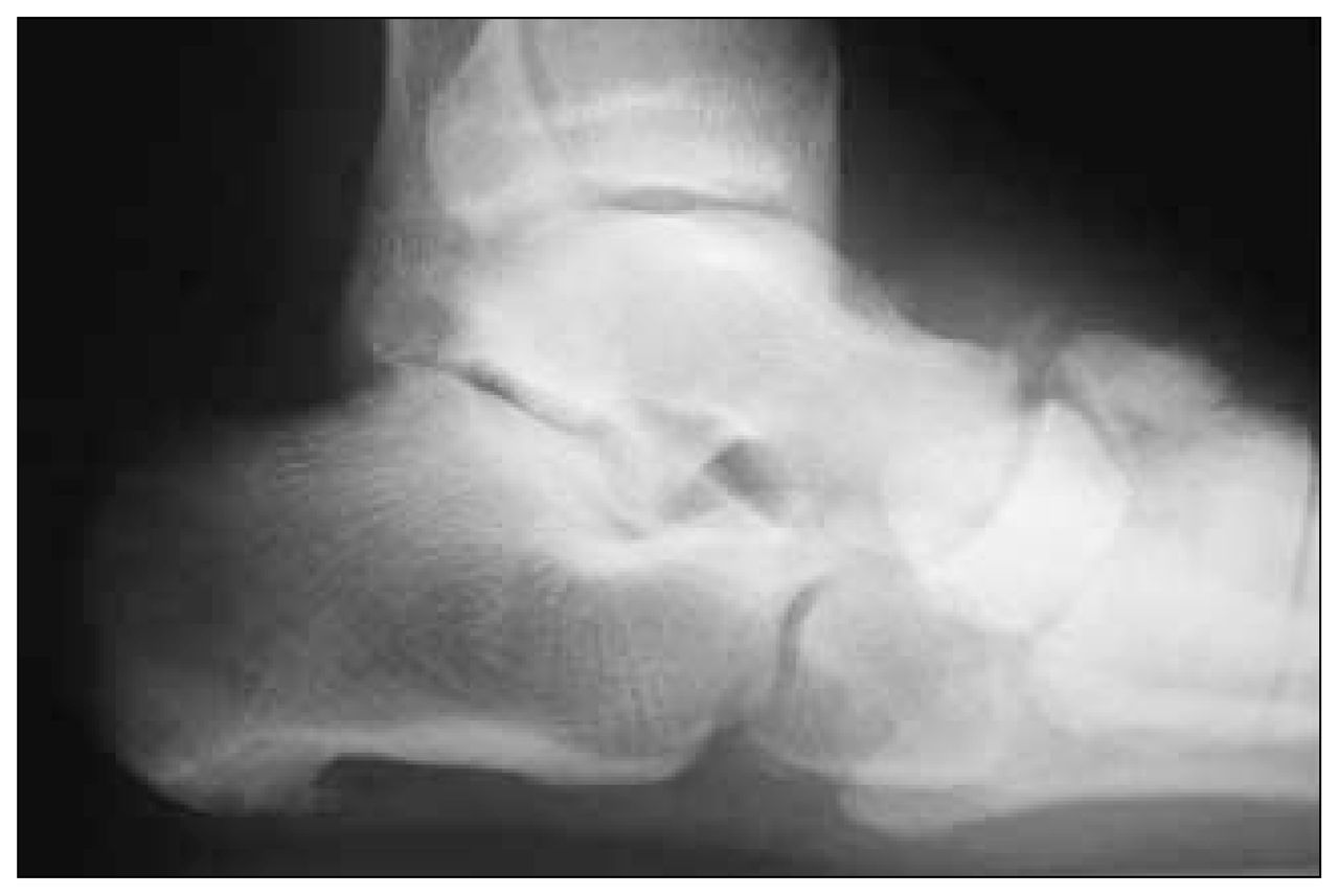
The patient returned 3 weeks later complaining of increased swelling, warmth and redness in the foot. She had no particular complaints of pain. On this occasion the family physician obtained a radiograph (Fig. 1).

FIG. 1
  • Download figure
  • Open in new tab
  • Download powerpoint
FIG. 1

Because of concern over the possibility of infection secondary to her known diabetes, the patient was referred for needle aspiration at the hospital clinic. No purulent material was identified, and the fluid specimen obtained was heavily blood stained. Cultures of the specimen produced no growth.

Eventually the patient was referred for orthopedic assessment. At that time she noted significant deformity of the foot with increasing difficulty walking. The foot was red, swollen and deformed with increased fullness on the plantar surface and obvious abduction of the forefoot.

New radiographs (Fig. 2) were obtained. A diagnosis of Charcot arthropathy was made, based on the extent of disorganization of the foot, lack of significant pain, rapid progression of bone destruction and negative findings on culture of the aspiration specimen.

FIG. 2
  • Download figure
  • Open in new tab
  • Download powerpoint
FIG. 2

Charcot arthropathy is a well-recognized disorder primarily seen in the lower extremities of patients with impaired position sense or impaired pain perception. Common conditions associated with Charcot arthropathy include tabes dorsalis, diabetes mellitus and syringomyelia. Usually occurring in the weight-bearing joints of the lower extremity it can also be seen in the upper extremity usually in the shoulder or elbow. Typical findings in the upper extremity include relatively painless joint destruction usually associated with instability. Common symptoms in the lower extremity consist of moderate discomfort, increasing deformity and characteristic radiographic features including bone destruction, joint disorganization and atypical new bone formation.

Treatment for these patients varies according to their age, the joint involved and extent of joint disorganization and the presence of adjacent bone destruction.

Conventional orthopedic joint procedures such as arthrodesis or arthroplasty have a recognized higher failure rate in neuropathic joints, and conservative treatment, if possible, is recommended. With regard to the management of Charcot arthropathy of the foot, early recognition of the problem is the key to satisfactory management. If the foot can be supported appropriately to prevent excessive deformity a satisfactory outcome may be obtained. Significant deformity is, unfortunately, often associated with unacceptable points of pressure resulting in skin breakdown and ulceration, deep infection and eventually amputation (Fig. 3). Surgical realignment is recommended if pressure off-loading (bracing) is unsuccessful in healing the ulcer, in severe deformities or when a deformity progresses in spite of full-time bracing. The surgical goal is to provide the patient with a braceable extremity that is ulcer free. The use of rigid internal fixation has improved the outcomes, making surgical realignment a viable alternative to amputation of the foot.

FIG. 3
  • Download figure
  • Open in new tab
  • Download powerpoint
FIG. 3

Footnotes

  • Submissions to Surgical Images, musculoskeletal section, should be sent to the section editor: Dr. Norman S. Schachar, Heritage Medical Research Building, 36–3330 Hospital Dr. NW, Calgary AB T2N 4N1; fax 403 270-0617.

PreviousNext
Back to top

In this issue

Canadian Journal of Surgery: 45 (5)
CAN J SURG
Vol. 45, Issue 5
1 Oct 2002
  • Table of Contents
  • Index by author

Article tools

Respond to this article
Print
Download PDF
Article Alerts
To sign up for email alerts or to access your current email alerts, enter your email address below:
Email Article

Thank you for your interest in spreading the word on CJS.

NOTE: We only request your email address so that the person you are recommending the page to knows that you wanted them to see it, and that it is not junk mail. We do not capture any email address.

Enter multiple addresses on separate lines or separate them with commas.
Musculoskeletal images. Charcot arthropathy
(Your Name) has sent you a message from CJS
(Your Name) thought you would like to see the CJS web site.
CAPTCHA
This question is for testing whether or not you are a human visitor and to prevent automated spam submissions.
Citation Tools
Musculoskeletal images. Charcot arthropathy
Timothy Daniels, James P. Waddell
CAN J SURG Oct 2002, 45 (5) 363-364;

Citation Manager Formats

  • BibTeX
  • Bookends
  • EasyBib
  • EndNote (tagged)
  • EndNote 8 (xml)
  • Medlars
  • Mendeley
  • Papers
  • RefWorks Tagged
  • Ref Manager
  • RIS
  • Zotero
‍ Request Permissions
Share
Musculoskeletal images. Charcot arthropathy
Timothy Daniels, James P. Waddell
CAN J SURG Oct 2002, 45 (5) 363-364;
Digg logo Reddit logo Twitter logo Facebook logo Google logo Mendeley logo
  • Tweet Widget
  • Facebook Like

Related Articles

  • No related articles found.
  • PubMed
  • Google Scholar

Cited By...

  • No citing articles found.
  • Google Scholar

Similar Articles

Collections

  • Surgical Images

Content

  • Current issue
  • Past issues
  • Collections
  • Alerts
  • RSS

Authors & Reviewers

  • Overview for Authors
  • Publication Fees
  • Forms
  • Editorial Policies
  • Submit a manuscript

About

  • General Information
  • Staff
  • Editorial Board
  • Contact Us
  • Advertising
  • Reprints
  • Copyright and Permissions
  • Accessibility
  • CMA Civility Standards
CMAJ Group

Copyright 2023, CMA Impact Inc. or its licensors. All rights reserved. ISSN 2291-0026

All editorial matter in CJS represents the opinions of the authors and not necessarily those of the Canadian Medical Association or its subsidiaries.

To receive any of these resources in an accessible format, please contact us at CMAJ Group, 500-1410 Blair Towers Place, Ottawa ON, K1J 9B9; p: 1-888-855-2555; e: [email protected].

View CMA's Accessibility policy.

Powered by HighWire