
Main menu

  • Home
  • Content
    • Current issue
    • Past issues
    • Sections
    • Collections
  • Podcasts
  • Author Info
    • Overview for authors
    • Publication fees
    • Forms
    • Editorial policies
    • Submit a manuscript
    • Open access
  • Careers
  • Alerts
    • Email alerts
    • RSS
  • About
    • General information
    • Staff
    • Editorial Board
    • Contact
  • CMAJ JOURNALS
    • CMAJ
    • CMAJ Open
    • JAMC
    • JPN

User menu

Search

  • Advanced search
CJS
  • CMAJ JOURNALS
    • CMAJ
    • CMAJ Open
    • JAMC
    • JPN
CJS

Advanced Search

  • Home
  • Content
    • Current issue
    • Past issues
    • Sections
    • Collections
  • Podcasts
  • Author Info
    • Overview for authors
    • Publication fees
    • Forms
    • Editorial policies
    • Submit a manuscript
    • Open access
  • Careers
  • Alerts
    • Email alerts
    • RSS
  • About
    • General information
    • Staff
    • Editorial Board
    • Contact
  • Subscribe to our alerts
  • RSS feeds
  • Follow CJS on Twitter
Surgical Images

Soft-tissue images. Diffuse cavernous rectal hemangioma

Dursun Bugra, Turker Bulut, Sumer Yamaner, Rasim Gencosmanoglu, Vahit Ozmen, Orhan Bilge and Ugur Cevikbas
CAN J SURG October 01, 2002 45 (5) 365-366;
Dursun Bugra
*Department of General Surgery, University of Marmara, Istanbul, Turkey
  • Find this author on Google Scholar
  • Find this author on PubMed
  • Search for this author on this site
Turker Bulut
*Department of General Surgery, University of Marmara, Istanbul, Turkey
  • Find this author on Google Scholar
  • Find this author on PubMed
  • Search for this author on this site
Sumer Yamaner
*Department of General Surgery, University of Marmara, Istanbul, Turkey
  • Find this author on Google Scholar
  • Find this author on PubMed
  • Search for this author on this site
Rasim Gencosmanoglu
†Department of Surgical Unit, Institute of Gastroenterology, University of Marmara, Istanbul, Turkey
  • Find this author on Google Scholar
  • Find this author on PubMed
  • Search for this author on this site
Vahit Ozmen
*Department of General Surgery, University of Marmara, Istanbul, Turkey
  • Find this author on Google Scholar
  • Find this author on PubMed
  • Search for this author on this site
Orhan Bilge
*Department of General Surgery, University of Marmara, Istanbul, Turkey
  • Find this author on Google Scholar
  • Find this author on PubMed
  • Search for this author on this site
Ugur Cevikbas
‡Department of Pathology, Istanbul Medical Faculty, University of Istanbul
  • Find this author on Google Scholar
  • Find this author on PubMed
  • Search for this author on this site
  • Article
  • Figures & Tables
  • Responses
  • Metrics
  • PDF
Loading

A 27-year-old man, who had undergone hemorrhoidectomy 7 years previously, presented with intermittent rectal bleeding and anemia that had been present for 8 years. Digital rectal examination confirmed the rectal bleeding, but no mass was palpable in the rectal wall. The physical examination revealed no other abnormalities. Blood count showed mild anemia (hematocrit 27%, hemoglobin 79 g/L). Upper gastrointestinal endoscopy show no abnormalities, but colonoscopy revealed an edematous mucosa and diffuse submucosal bluish nodulation with dilated veins (Fig. 1), extending from the dentate line up for 20 cm. At laparotomy, the rectum and distal sigmoid colon had significant vascular engorgement on the serosal surface, extending through the mesorectum and mesosigmoid. The patient underwent low anterior resection with a stapled colorectal anastomosis. Diffuse cavernous hemangioma (DCH) was diagnosed.

FIG. 1
  • Download figure
  • Open in new tab
  • Download powerpoint
FIG. 1

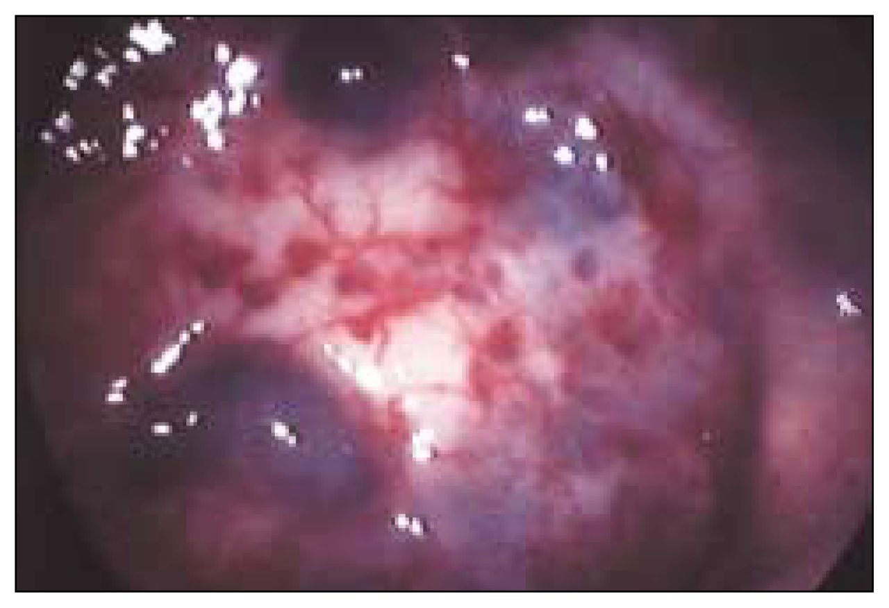
Endoscopic view of the diffuse cavernous hemangioma of the rectum.

The macroscopic view of the rectosigmoid colon showed many abnormal hemangiomatous vessels (Fig. 2). Histopathological examination of the specimen showed DCH invading all layers of rectum and distal sigmoid colon (Fig. 3). The patient was free of any symptoms at 6-year follow-up.

FIG. 2
  • Download figure
  • Open in new tab
  • Download powerpoint
FIG. 2

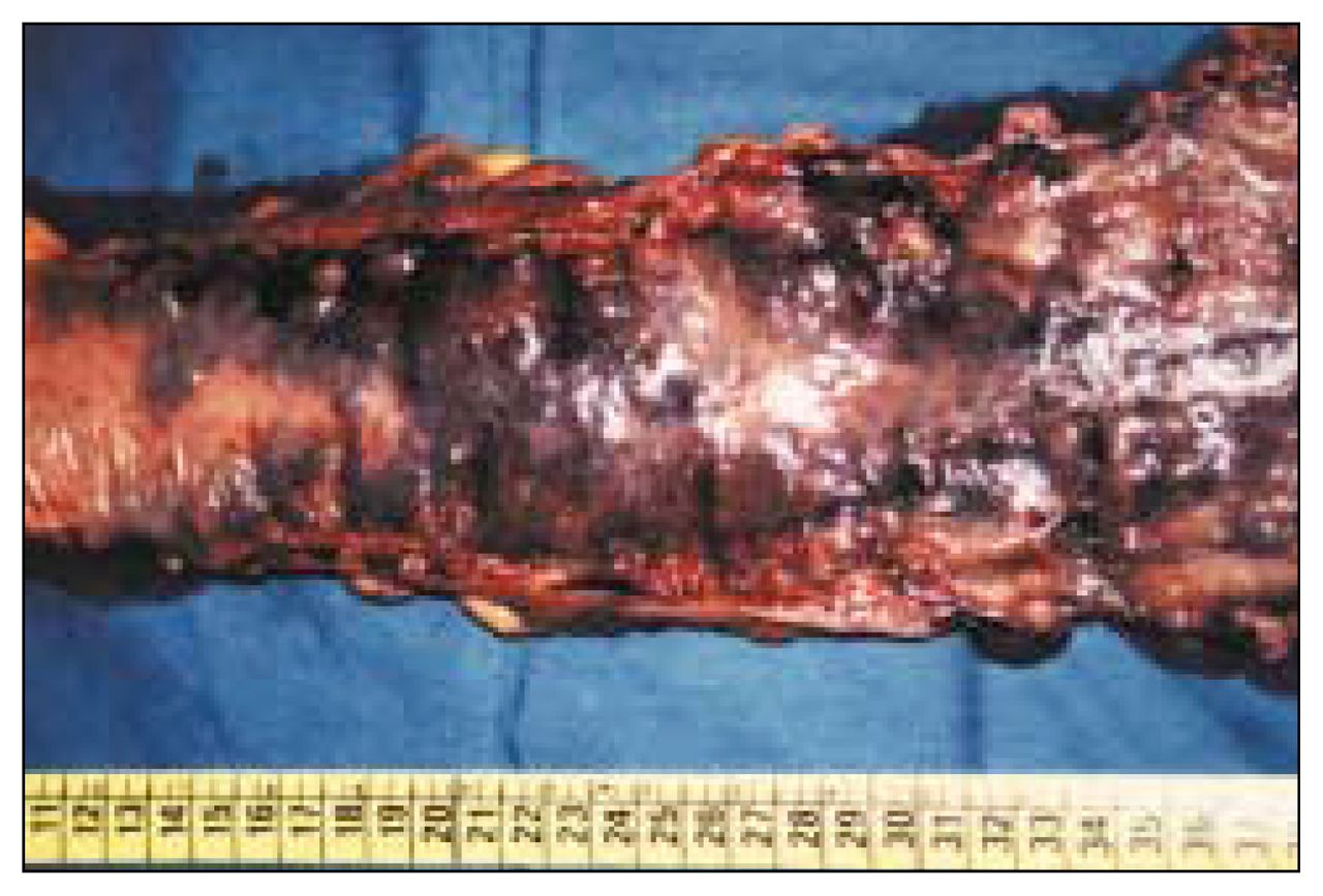
Macroscopic view of the excised rectal cavernous hemangioma.

FIG. 3
  • Download figure
  • Open in new tab
  • Download powerpoint
FIG. 3

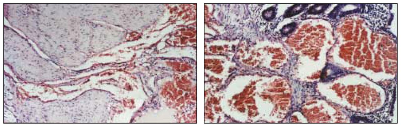
Microscopic findings of the cavernous hemangioma of the rectum show (a) extremely dilated vessels containing erythrocytes in all the layers of rectal wall and (b) the absence of cellular dysplasia in these vessels and the adjacent tissue, proving its hamartomatous nature (hematoxylin–eosin stain, original magnification ×350).

DCH of the rectosigmoid colon is an extremely rare malformation that is generally seen in young adults, with a male-to-female ratio of 2:1.1 Patients usually suffer painless rectal bleeding and anemia. However, there may be associated intestinal obstruction, diarrhea, constipation and hematuria.2 The presence of hemangioma in the cutaneous and mucous membranes, or Klippel–Trenaunay syndrome, should alert the clinician to the presence of rectal hemangioma.3,4 Endoscopy is the method of choice for diagnosis. Upper gastrointestinal endoscopy should be done in all patients to rule out any synchronous hemangiomas. Other diagnostic tools such as barium enema, selective angiography, computed tomography, magnetic resonance imaging or endoscopic ultrasonography may show extramural spread of the lesion and exclude other causes of rectal bleeding. The definitive treatment for DCH is excision of the involved bowel. Sphincter-saving procedures, avoiding damage to perirectal neural structures, is preferred for satisfactory long-term results.5 The vascular malformations that remain in the proximal anal canal, usually 1 to 2 cm above the dentate line, can be managed conservatively by argon or Nd:YAG laser therapy, or sclerotherapy in the case of rebleeding.

Footnotes

  • Submissions to Surgical Images, soft-tissue section, should be sent to the section editors: Dr. David P. Girvan, Victoria Hospital Corporation, PO Box 5375, Station B, London ON N6A 5A5 or Dr. Nis Schmidt, Department of Surgery, St. Paul’s Hospital, 1081 Burrard St., Vancouver BC V6Z 1Y6.

References

  1. ↵
    1. Cunningham JA,
    2. Garcia VF,
    3. Quispe G
    . Diffuse cavernous rectal hemangioma — sphincter-sparing approach to therapy. Report of a case. Dis Colon Rectum 1989; 32:344–7.
    OpenUrlPubMed
  2. ↵
    1. Demircan O,
    2. Sonmez H,
    3. Zeren S,
    4. Cosar E,
    5. Bicakci K,
    6. Ozkan S
    . Diffuse cavernous hemangioma of the rectum and sigmoid colon. Dig Surg 1998;15:713–5.
    OpenUrlCrossRefPubMed
  3. ↵
    1. Hagood MF,
    2. Gathright JB Jr.
    . Hemangiomatosis of the skin and the gastrointestinal tract: report of a case. Dis Colon Rectum 1975;18:141–6.
    OpenUrlCrossRefPubMed
  4. ↵
    1. Azizkhan EG
    . Life threatening hematochezia from a rectosigmoid vascular malformation in Klippel–Trenaunay syndrome: long term palliation using an argon laser. J Pediatr Surg 1991;26:1125–7.
    OpenUrlPubMed
  5. ↵
    1. Oner Z,
    2. Altaca G
    . Diffuse cavernous rectal hemangioma — clinical appearance, diagnostic modalities and sphincter saving approach to therapy: report of 2 and a collective review of 79 cases [review]. Acta Chir Belg 1993;93:173–6.
    OpenUrlPubMed
PreviousNext
Back to top

In this issue

Canadian Journal of Surgery: 45 (5)
CAN J SURG
Vol. 45, Issue 5
1 Oct 2002
  • Table of Contents
  • Index by author

Article tools

Respond to this article
Print
Download PDF
Article Alerts
To sign up for email alerts or to access your current email alerts, enter your email address below:
Email Article

Thank you for your interest in spreading the word on CJS.

NOTE: We only request your email address so that the person you are recommending the page to knows that you wanted them to see it, and that it is not junk mail. We do not capture any email address.

Enter multiple addresses on separate lines or separate them with commas.
Soft-tissue images. Diffuse cavernous rectal hemangioma
(Your Name) has sent you a message from CJS
(Your Name) thought you would like to see the CJS web site.
CAPTCHA
This question is for testing whether or not you are a human visitor and to prevent automated spam submissions.
Citation Tools
Soft-tissue images. Diffuse cavernous rectal hemangioma
Dursun Bugra, Turker Bulut, Sumer Yamaner, Rasim Gencosmanoglu, Vahit Ozmen, Orhan Bilge, Ugur Cevikbas
CAN J SURG Oct 2002, 45 (5) 365-366;

Citation Manager Formats

  • BibTeX
  • Bookends
  • EasyBib
  • EndNote (tagged)
  • EndNote 8 (xml)
  • Medlars
  • Mendeley
  • Papers
  • RefWorks Tagged
  • Ref Manager
  • RIS
  • Zotero
‍ Request Permissions
Share
Soft-tissue images. Diffuse cavernous rectal hemangioma
Dursun Bugra, Turker Bulut, Sumer Yamaner, Rasim Gencosmanoglu, Vahit Ozmen, Orhan Bilge, Ugur Cevikbas
CAN J SURG Oct 2002, 45 (5) 365-366;
Digg logo Reddit logo Twitter logo Facebook logo Google logo Mendeley logo
  • Tweet Widget
  • Facebook Like

Related Articles

  • No related articles found.
  • PubMed
  • Google Scholar

Cited By...

  • No citing articles found.
  • Google Scholar

Similar Articles

Collections

  • Surgical Images

Content

  • Current issue
  • Past issues
  • Collections
  • Alerts
  • RSS

Authors & Reviewers

  • Overview for Authors
  • Publication Fees
  • Forms
  • Editorial Policies
  • Submit a manuscript

About

  • General Information
  • Staff
  • Editorial Board
  • Contact Us
  • Advertising
  • Reprints
  • Copyright and Permissions
  • Accessibility
  • CMA Civility Standards
CMAJ Group

Copyright 2023, CMA Impact Inc. or its licensors. All rights reserved. ISSN 2291-0026

All editorial matter in CJS represents the opinions of the authors and not necessarily those of the Canadian Medical Association or its subsidiaries.

To receive any of these resources in an accessible format, please contact us at CMAJ Group, 500-1410 Blair Towers Place, Ottawa ON, K1J 9B9; p: 1-888-855-2555; e: [email protected].

View CMA's Accessibility policy.

Powered by HighWire