
Main menu

  • Home
  • Content
    • Current issue
    • Past issues
    • Sections
    • Collections
  • Podcasts
  • Author Info
    • Overview for authors
    • Publication fees
    • Forms
    • Editorial policies
    • Submit a manuscript
    • Open access
  • Careers
  • Alerts
    • Email alerts
    • RSS
  • About
    • General information
    • Staff
    • Editorial Board
    • Contact
  • CMAJ JOURNALS
    • CMAJ
    • CMAJ Open
    • JAMC
    • JPN

User menu

Search

  • Advanced search
CJS
  • CMAJ JOURNALS
    • CMAJ
    • CMAJ Open
    • JAMC
    • JPN
CJS

Advanced Search

  • Home
  • Content
    • Current issue
    • Past issues
    • Sections
    • Collections
  • Podcasts
  • Author Info
    • Overview for authors
    • Publication fees
    • Forms
    • Editorial policies
    • Submit a manuscript
    • Open access
  • Careers
  • Alerts
    • Email alerts
    • RSS
  • About
    • General information
    • Staff
    • Editorial Board
    • Contact
  • Subscribe to our alerts
  • RSS feeds
  • Follow CJS on Twitter
Radiology for the Surgeon

Musculoskeletal case 24. Diagnosis

CAN J SURG October 01, 2002 45 (5) 389-390;
  • Article
  • Figures & Tables
  • Responses
  • Metrics
  • PDF
Loading

Intramuscular lipoma

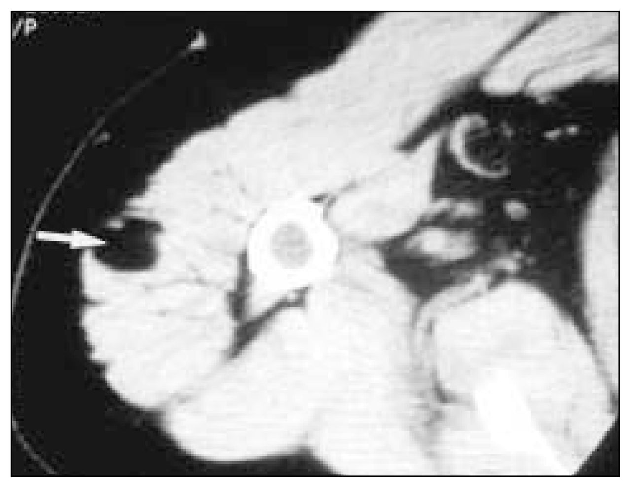
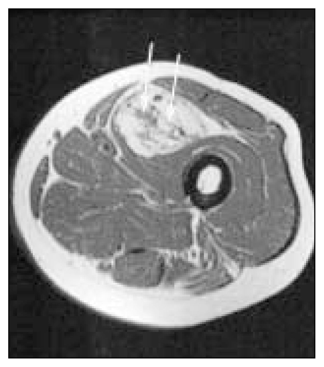
The axial computed tomography scan (Fig. 1) revealed a hypodense, well-defined lesion (arrow) within the deltoid muscle at the level of the humeral neck. The lesion was similar in attenuation to adjacent subcutaneous fat.

FIG. 1
  • Download figure
  • Open in new tab
  • Download powerpoint
FIG. 1

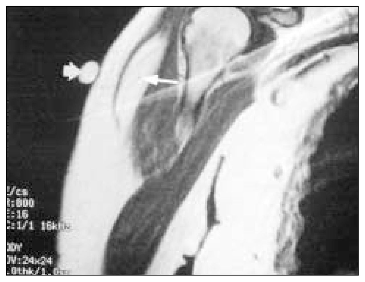
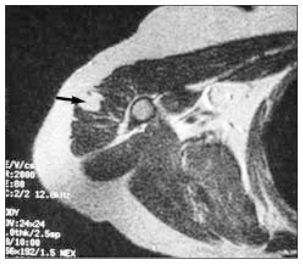
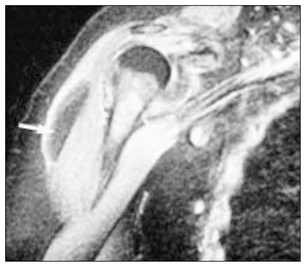
The T1-weighted coronal oblique magnetic resonance image of the area (Fig. 2), obtained for further assessment of the lump, demonstrated a well-defined hyperintense area (arrow) within the deltoid muscle, of similar signal intensity to adjacent fat. The hyperintense nodule (arrowhead) on the skin is a vitamin E capsule used to mark the area of interest. A fat-soluble vitamin, it has the same signal characteristics as fat within the body. On axial T2-weighted (Fig. 3) and proton density-weighted (not shown) images, the lesion (arrow) again had similar signal characteristics to fat. A fat-suppressed coronal oblique scan demonstrated suppression of the signal from the abnormality (Fig. 4, arrow).

FIG. 2
  • Download figure
  • Open in new tab
  • Download powerpoint
FIG. 2
FIG. 3
  • Download figure
  • Open in new tab
  • Download powerpoint
FIG. 3
FIG. 4
  • Download figure
  • Open in new tab
  • Download powerpoint
FIG. 4

Lipomas, the most common benign soft-tissue neoplasms, can occur in any soft tissue that contains fat, including subcutaneous tissue, muscle, nerve and synovium, or adjacent to bone in the periosteum.1,2 People of any age group can be affected, although lipomas are more common in adults, with a slight male preponderance.3 Lipomas in muscle may be intermuscular or intramuscular and may not be recognized clinically unless they are large or symptomatic. Dysfunction of the muscle or pain (lipoma dolorosa) is rare.

If the lipoma is large enough, plain radiographs may reveal a relatively radiolucent lesion within the muscle, but computed tomography and magnetic resonance imaging scans better demonstrate the lesion. Lipomas should approximate to fat on all imaging sequences. In general they appear as homogeneous, sharply defined fatty masses.1,3 Septations, which may be thin fibrous bands or thicker muscle bundles, are not uncommon in intramuscular lipomas but may make it impossible to rule out well-differentiated liposarcoma on the basis of imaging alone.4 Fig. 5 is a T1-weighted MR image of a different patient with a biopsy-proven, moderately well-differentiated liposarcoma. Note that the lesion’s signal is predominantly of fat, but it has areas of banding and thick septations within.

FIG. 5
  • Download figure
  • Open in new tab
  • Download powerpoint
FIG. 5

Intramuscular myxoma and peripheral nerve sheath tumours may present as low-density masses on CT and simulate intramuscular lipoma.3 The measured CT attenuation values are usually not quite as low as those of subcutaneous fat (< –80 Hounsfield units). MR imaging can readily distinguish these lesions from lipoma. If doubt persists on imaging, surgical excision is required.

References

  1. ↵
    1. Resnick D
    . Diagnosis of bone and joint disorders. Philadelphia: W.B. Saunders; 1995. p. 3941–7.
  2. ↵
    1. Munk PL,
    2. Lee MJ,
    3. Janzen DL,
    4. Connell DG,
    5. Logan PM,
    6. Poon PY,
    7. et al
    . Lipoma and liposarcoma: evaluation using CT and MR imaging. AJR Am J Roentgenol 1997;169:589–94.
    OpenUrlPubMed
  3. ↵
    1. Kransdorf MJ,
    2. Moser RP Jr.,
    3. Meis MJ,
    4. Meyer CA
    . Fat-containing soft-tissue masses of the extremities. Radiographics 1991;11:81–106.
    OpenUrlCrossRefPubMed
  4. ↵
    1. Matsumoto K,
    2. Hukuda S,
    3. Ishizawa M,
    4. Chano T,
    5. Okabe H
    . MRI findings in intramuscular lipomas. Skeletal Radiol 1999;28:145–52.
    OpenUrlCrossRefPubMed
PreviousNext
Back to top

In this issue

Canadian Journal of Surgery: 45 (5)
CAN J SURG
Vol. 45, Issue 5
1 Oct 2002
  • Table of Contents
  • Index by author

Article tools

Respond to this article
Print
Download PDF
Article Alerts
To sign up for email alerts or to access your current email alerts, enter your email address below:
Email Article

Thank you for your interest in spreading the word on CJS.

NOTE: We only request your email address so that the person you are recommending the page to knows that you wanted them to see it, and that it is not junk mail. We do not capture any email address.

Enter multiple addresses on separate lines or separate them with commas.
Musculoskeletal case 24. Diagnosis
(Your Name) has sent you a message from CJS
(Your Name) thought you would like to see the CJS web site.
CAPTCHA
This question is for testing whether or not you are a human visitor and to prevent automated spam submissions.
Citation Tools
Musculoskeletal case 24. Diagnosis
CAN J SURG Oct 2002, 45 (5) 389-390;

Citation Manager Formats

  • BibTeX
  • Bookends
  • EasyBib
  • EndNote (tagged)
  • EndNote 8 (xml)
  • Medlars
  • Mendeley
  • Papers
  • RefWorks Tagged
  • Ref Manager
  • RIS
  • Zotero
‍ Request Permissions
Share
Musculoskeletal case 24. Diagnosis
CAN J SURG Oct 2002, 45 (5) 389-390;
Digg logo Reddit logo Twitter logo Facebook logo Google logo Mendeley logo
  • Tweet Widget
  • Facebook Like

Related Articles

  • No related articles found.
  • Google Scholar

Cited By...

  • No citing articles found.
  • Google Scholar

Similar Articles

Content

  • Current issue
  • Past issues
  • Collections
  • Alerts
  • RSS

Authors & Reviewers

  • Overview for Authors
  • Publication Fees
  • Forms
  • Editorial Policies
  • Submit a manuscript

About

  • General Information
  • Staff
  • Editorial Board
  • Contact Us
  • Advertising
  • Reprints
  • Copyright and Permissions
  • Accessibility
  • CMA Civility Standards
CMAJ Group

Copyright 2023, CMA Impact Inc. or its licensors. All rights reserved. ISSN 2291-0026

All editorial matter in CJS represents the opinions of the authors and not necessarily those of the Canadian Medical Association or its subsidiaries.

To receive any of these resources in an accessible format, please contact us at CMAJ Group, 500-1410 Blair Towers Place, Ottawa ON, K1J 9B9; p: 1-888-855-2555; e: [email protected].

View CMA's Accessibility policy.

Powered by HighWire