


A 65-year-old woman who presented with hematemesis and melena had a history of chronic anemia but no previous episodes of gastrointestinal bleeding. Her hemoglobin level was 69 g/L. Gastroscopy revealed a submucosal mass in the posterior wall of the antrum and focal ulcerations of the mucosa overlying the lesion. An upper gastrointestinal series confirmed a round, well-circumscribed mass in the antrum (Fig. 1).

Upper gastrointestinal series demonstrates a gastrointestinal stromal tumour (GIST) of the antrum.
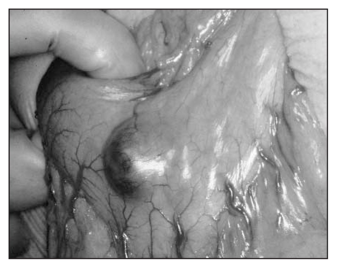
At laparotomy, the mass could be seen impinging on the anterior gastric wall (Fig. 2). A distal gastrectomy was performed. Examination of the gross specimen revealed a 3.5 × 2.5 × 2.2 cm submucosal polypoid mass and erosions of the mucosa (Fig. 3).

The GIST is impinging on the anterior gastric wall.
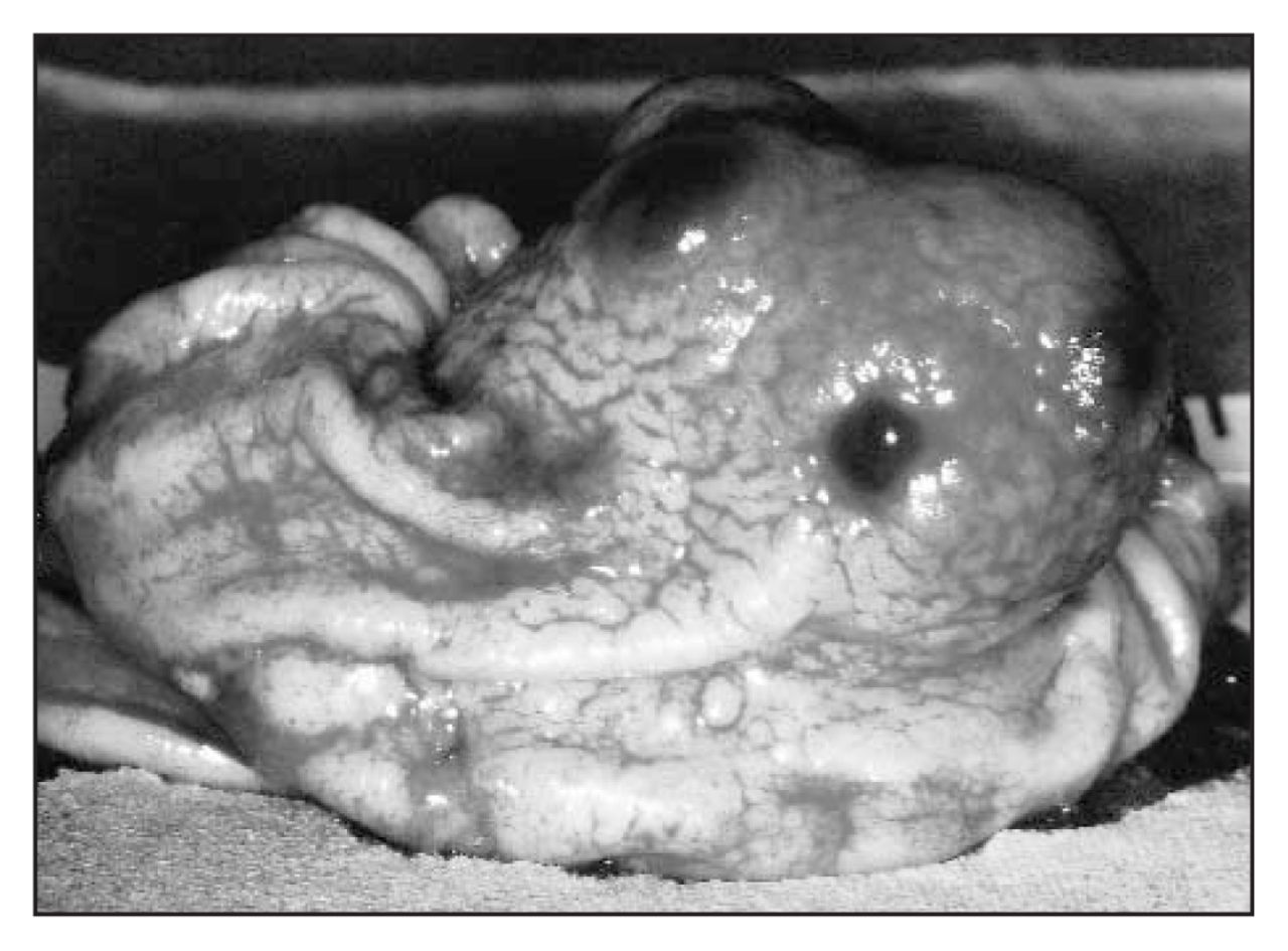
The GIST specimen showing a submucosal polypoid mass and erosions of the mucosa.
Histologic analysis showed a gastrointestinal stromal tumour (GIST), spindle cell type, composed of eosinophilic cells arranged in short fascicles and a paler syncytial-appearing eosinophilic cytoplasm (Fig. 4).

Histologically, the GIST is of the spindle cell type (hematoxylin–eosin stain; original magnification ×400).
Mitotic activity did not exceed 2 mitoses per 50 high-power fields (HPFs). The tumour was immunochemically slightly positive for muscle-specific actin, and negative for desmin S-100. Immunostaining for the KIT receptor was not performed because this technique has just been implemented at our institution. The tumour was classified as benign based on its size (<5 cm) and according to its negligible mitotic activity.
GIST describes the largest category of primary nonepithelial nonlymphomatous neoplasms of the stomach and small bowel. Gastric stromal tumours are rare, accounting for only about 4% of all the tumours of the stomach. Clinical outcome and prognosis are difficult to predict from morphologic data. These tumours are characterized by a remarkable cellular variability, and their malignant potential is sometimes difficult to predict. GISTs are usually categorized into 3 groups: (1) benign: tumour size less than 5 cm, mitotic count less than 5 per 50 HPFs; (2) borderline: tumour size more than 5 cm and less than 10 cm, mitotic count no more than 5 per 50 HPFs; (3) malignant: tumour any size, mitotic count greater than 5 per 50 HPFs.1–3
Although modern immunohistochemical techniques are available, mitotic count is one of the more reliable single factors in differentiating between GISTs of varying malignant potential.4
Footnotes
Submissions to Surgical Images, soft-tissue section, should be sent to the section editors: Dr. David P. Girvan, Victoria Hospital Corporation, PO Box 5375, Station B, London ON N6A 5A5 or Dr. Nis Schmidt, Department of Surgery, St. Paul’s Hospital, 1081 Burrard St., Vancouver BC V6Z 1Y6.
Competing interests: None declared.
In this issue

Article tools


Related Articles
Cited By...
- No citing articles found.