
Main menu

  • Home
  • Content
    • Current issue
    • Past issues
    • Sections
    • Collections
  • Podcasts
  • Author Info
    • Overview for authors
    • Publication fees
    • Forms
    • Editorial policies
    • Submit a manuscript
    • Open access
  • Careers
  • Alerts
    • Email alerts
    • RSS
  • About
    • General information
    • Staff
    • Editorial Board
    • Contact
  • CMAJ JOURNALS
    • CMAJ
    • CMAJ Open
    • JAMC
    • JPN

User menu

Search

  • Advanced search
CJS
  • CMAJ JOURNALS
    • CMAJ
    • CMAJ Open
    • JAMC
    • JPN
CJS

Advanced Search

  • Home
  • Content
    • Current issue
    • Past issues
    • Sections
    • Collections
  • Podcasts
  • Author Info
    • Overview for authors
    • Publication fees
    • Forms
    • Editorial policies
    • Submit a manuscript
    • Open access
  • Careers
  • Alerts
    • Email alerts
    • RSS
  • About
    • General information
    • Staff
    • Editorial Board
    • Contact
  • Subscribe to our alerts
  • RSS feeds
  • Follow CJS on Twitter
Continuing Medical Education

Calcinosis cutis

Luc A. Dubois, Daryl K. Gray and Edward J. Tweedie
Can J Surg June 01, 2007 50 (3) 217-218;
Luc A. Dubois
*Division of General Surgery, University of Western Ontario, London, Ont.
MD
  • Find this author on Google Scholar
  • Find this author on PubMed
  • Search for this author on this site
Daryl K. Gray
*Division of General Surgery, University of Western Ontario, London, Ont.
MD
  • Find this author on Google Scholar
  • Find this author on PubMed
  • Search for this author on this site
Edward J. Tweedie
†Division of Pathology, University of Western Ontario, London, Ont.
MD
  • Find this author on Google Scholar
  • Find this author on PubMed
  • Search for this author on this site
  • Article
  • Figures & Tables
  • Responses
  • Metrics
  • PDF
Loading

A 71-year-old woman with new onset end-stage renal failure of unknown origin presented with symptoms of uremia (nausea, vomiting, purities, dyspnea, fatigue). Physical examination revealed an area of dry gangrene on the left leg. Laboratory investigations demonstrated a urea of 47.6 mmol/L, creatinine level of 884 μmol/L, hemoglobin of 60 g/L, calcium of 3.5 mmol/L, a phosphate level of 2.15 mmol/L, a calcium phosphorus product of 93 ([Ca] × [PO3] in conventional units) and a parathyroid hormone of 1138 pg/mL. The patient was placed on hemodialysis. The gangrenous lesion was presumed to be related to the patient’s venous insufficiency.

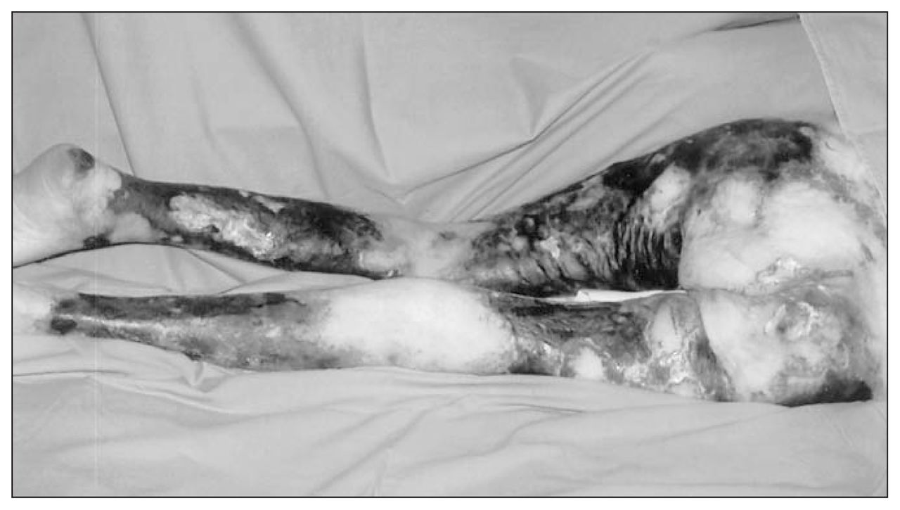
Over the next 2 months, there was progression of the dry gangrene involving the anterior and posterior aspect of both upper legs, the posterior and lateral aspect of the lower legs, the lower back and the buttocks (Fig. 1). A thromboembolic etiology was considered, and the patient was placed on heparin. A skin biopsy of the lesion showed no evidence of emboli, and an echocardiogram failed to reveal a source.

FIG. 1
  • Download figure
  • Open in new tab
  • Download powerpoint
FIG. 1

Photograph taken at operating room showing the extent of the necrotic skin lesions.

The patient was referred to our centre, where a bilateral leg x-ray revealed calcification of the small arteries of both legs (Fig. 2). A second skin biopsy established the diagnosis of calcinosis cutis (Fig. 3) due to hyperparathyroidism. An urgent parathyroidectomy was performed. At parathyroidectomy, a large, fixed, right superior parathyroid gland and a normal looking right inferior gland were visualized and removed.

FIG. 2
  • Download figure
  • Open in new tab
  • Download powerpoint
FIG. 2

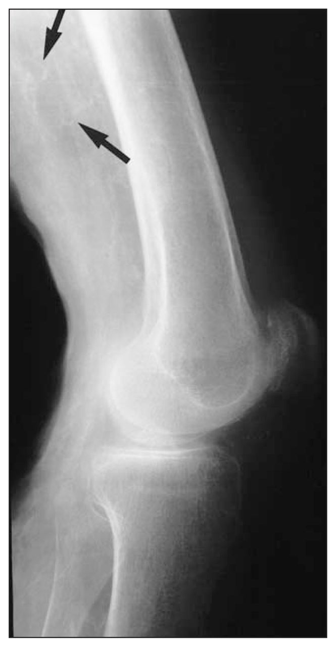
Radiograph of the left leg. The arrow shows calcified small blood vessels.

FIG. 3
  • Download figure
  • Open in new tab
  • Download powerpoint
FIG. 3

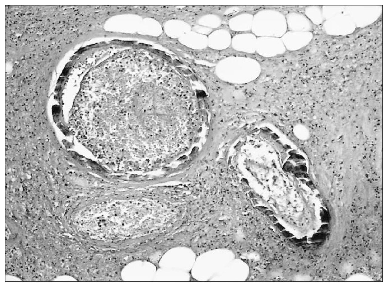
100 × (hematoxylin and eosin) micrograph of cutaneous arteries showing extensive mural calcification, luminal thrombosis and necrosis of surrounding tissue.

On gross examination, the right superior specimen weighed 3.96 g, with the gland measuring from 2.7 × 2.1 × 1.5 cm in size (Fig. 4). The inferior parathyroid gland measured 1 × 0.65 × 0.4 cm and weighed 0.073 g. Microscopic examination of the right superior gland demonstrated hypercellularity and calcification. Gross and microscopic findings were consistent with parathyroid adenoma.

FIG. 4
  • Download figure
  • Open in new tab
  • Download powerpoint
FIG. 4

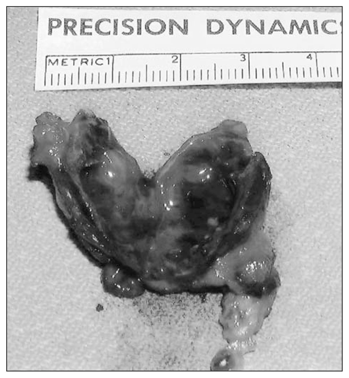
Right superior parathyroid adenoma (3.96 g).

The patient’s condition deteriorated postoperatively; she developed pneumonia and bilateral pleural effusions and became hypotensive. Due to the extent of the necrotic tissue in her legs, the need for surgical débridement and amputation, and her concomitant medical problems, the family decided to withdraw care. The patient died of overwhelming sepsis.

Calcinosis cutis (also known as calciphylaxis), the progressive deposition of calcium in the skin and small blood vessels with associated thrombosis and necrosis, is a rare and life-threatening illness. It most commonly results from hyperparathyroidism secondary to renal failure and most commonly occurs in women.1 Calcinosis cutis typically presents with ulcerating painful purplish lesions in the extremities and trunk. These lesions often progress to necrosis and act as a portal for infectious agents leading to wound sepsis and death.2 The etiology of this disease remains unclear, however, and elevated calcium phosphorous product ([Ca] × [PO3] > 70 in conventional units) is thought to act as a sensitizer, promoting calcification. Elevated levels of parathyroid hormone have also been implicated as a sensitizer.1 Treatment is difficult, with such medical therapies as phosphate binders and steroids having little effect.3 The best current therapy is systemic antibiotic drugs, débridement of necrotic tissue and semi-urgent parathyroidectomy.1–4 Early parathyroidectomy has been shown to significantly reduce mortality in a study of 104 patients with calcinosis cutis and renal failure.4 Despite these results, the literature would agree that the current therapy remains unsatisfactory. 1–3 Calcinosis cutis can be difficult to diagnose on pathology; thus, if the clinical suspicion of calcinosis remains high after a negative skin biopsy, it is wise to repeat it.

Footnotes

  • Submissions to the Surgical Images, soft-tissue section, should be sent to the section editors: Dr. David P. Girvan, Victoria Hospital Corporation, PO Box 5375, Station B, London ON N6A 5A5 or Dr. Nis Schmidt, Department of Surgery, St. Paul’s Hospital, 1081 Burrard St., Vancouver BC V6Z 1Y6.

  • Competing interests: None declared.

References

  1. ↵
    1. Ledbetter LS,
    2. Khoshnevis MR,
    3. Hsu S
    . Calciphylaxis. Cutis 2000;66:49–51.
    OpenUrlPubMed
  2. ↵
    1. Gilson RT
    . Calciphylaxis: case report and treatment review. Cutis 1999;63:149–53.
    OpenUrl
  3. ↵
    1. Shriner DL,
    2. Leevy CB,
    3. Schwartz RA,
    4. et al
    . Calcinosis cutis associated with secondary hyperparathyroidism due to renal failure. Int J Dermatol 1996;35:885–7.
    OpenUrlCrossRefPubMed
  4. ↵
    1. Hafner J,
    2. Keusch G,
    3. Wahl C,
    4. et al
    . Uremic small-artery disease with medical calcification and intimal hyperplasia (so-called caciphylaxis): a complication of chronic renal failure and benefit from parathyroidectomy. J Am Acad Dermatol 1995;33:954–62.
    OpenUrlCrossRefPubMed
PreviousNext
Back to top

In this issue

Canadian Journal of Surgery: 50 (3)
Can J Surg
Vol. 50, Issue 3
1 Jun 2007
  • Table of Contents
  • Table of Contents (PDF)
  • Index by author

Article tools

Respond to this article
Print
Download PDF
Article Alerts
To sign up for email alerts or to access your current email alerts, enter your email address below:
Email Article

Thank you for your interest in spreading the word on CJS.

NOTE: We only request your email address so that the person you are recommending the page to knows that you wanted them to see it, and that it is not junk mail. We do not capture any email address.

Enter multiple addresses on separate lines or separate them with commas.
Calcinosis cutis
(Your Name) has sent you a message from CJS
(Your Name) thought you would like to see the CJS web site.
CAPTCHA
This question is for testing whether or not you are a human visitor and to prevent automated spam submissions.
Citation Tools
Calcinosis cutis
Luc A. Dubois, Daryl K. Gray, Edward J. Tweedie
Can J Surg Jun 2007, 50 (3) 217-218;

Citation Manager Formats

  • BibTeX
  • Bookends
  • EasyBib
  • EndNote (tagged)
  • EndNote 8 (xml)
  • Medlars
  • Mendeley
  • Papers
  • RefWorks Tagged
  • Ref Manager
  • RIS
  • Zotero
‍ Request Permissions
Share
Calcinosis cutis
Luc A. Dubois, Daryl K. Gray, Edward J. Tweedie
Can J Surg Jun 2007, 50 (3) 217-218;
Digg logo Reddit logo Twitter logo Facebook logo Google logo Mendeley logo
  • Tweet Widget
  • Facebook Like

Related Articles

  • No related articles found.
  • PubMed
  • Google Scholar

Cited By...

  • No citing articles found.
  • Google Scholar

Similar Articles

Collections

  • Surgical Images

Content

  • Current issue
  • Past issues
  • Collections
  • Alerts
  • RSS

Authors & Reviewers

  • Overview for Authors
  • Publication Fees
  • Forms
  • Editorial Policies
  • Submit a manuscript

About

  • General Information
  • Staff
  • Editorial Board
  • Contact Us
  • Advertising
  • Reprints
  • Copyright and Permissions
CMAJ Group

Copyright 2023, CMA Impact Inc. or its licensors. All rights reserved. Print ISSN 0008-428X; Online ISSN 1488-2310.

All editorial matter in CJS represents the opinions of the authors and not necessarily those of the Canadian Medical Association or its subsidiaries.

To receive any of these resources in an accessible format, please contact us at CMAJ Group, 500-1410 Blair Towers Place, Ottawa ON, K1J 9B9; p: 1-888-855-2555; e: [email protected].

CMA Civility, Accessibility, Privacy


Powered by HighWire