


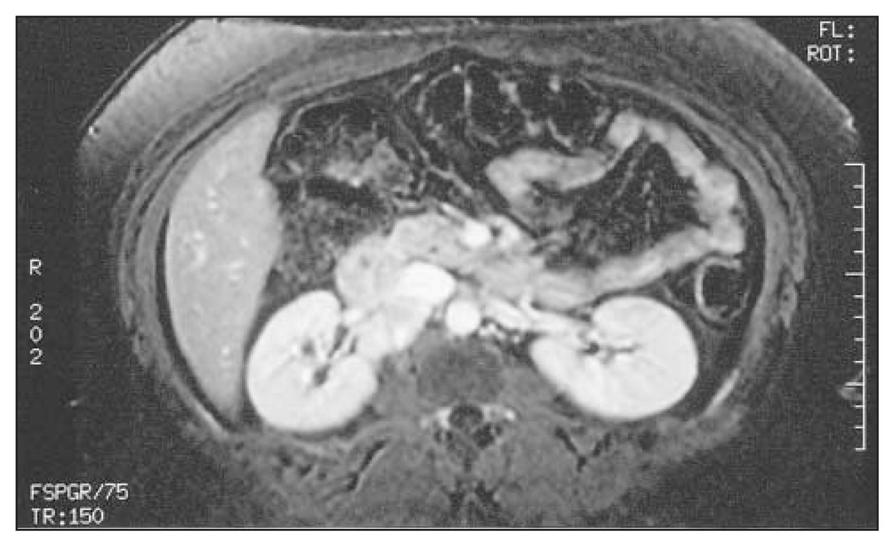
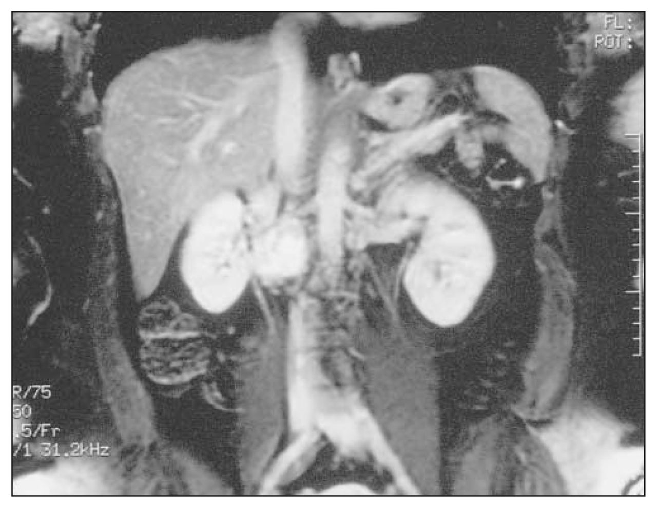
A 21-year-old woman, weighing 121 kg, presented with blurred vision. On examination, her blood pressure was 210/160 mm Hg. The physical examination was otherwise unremarkable. She was found to have elevated urinary dopamine, normetanephrine and total metanephrine levels. Magnetic resonance imaging (Fig. 1 and Fig. 2) demonstrated a dumbell-shaped tumour, measuring 4.0 × 3.5 × 2.6 cm, in the right renal hilum and surrounding the right renal vein. Both adrenal glands were normal. The patient underwent preoperative blockade with phenoxybenzamine, atenolol and metyrosine for 2 weeks.
Magnetic resonance image shows the dumbell-like tumour surrounding the right renal vein.

Magnetic resonance image shows the dumbell-like lesion in the region of the right renal hilum.
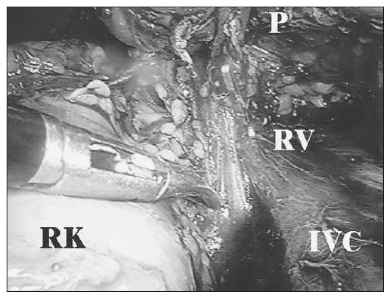
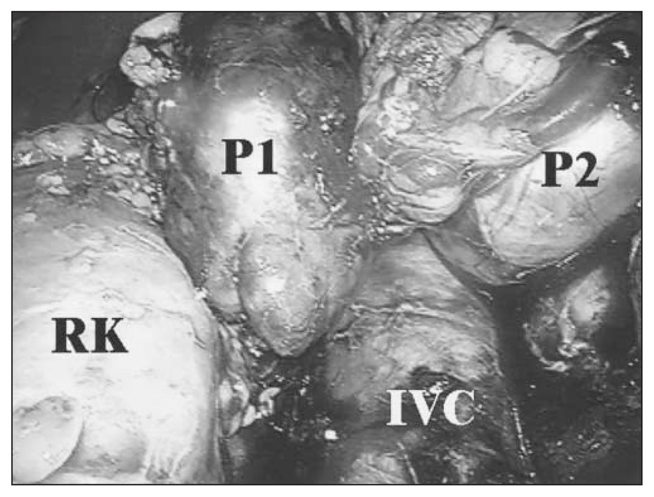
Through a transperitoneal laparoscopic approach, using 4 ports and with the patient in the right flank position, the hepatic flexure of the colon was mobilized and the duodenum reflected medially to expose the right renal hilum. After circumferential dissection of the lesion with use of the harmonic scalpel, 2 separate tumours with a fibrous connection were identified (Fig. 3). The tumours were adherent to the right renal vein but were successfully resected intact (Fig. 4). The estimated blood loss was 75 mL, and the operating time was 245 minutes. Pathological examination demonstrated that both tumours were pheochromocytomas and the surgical margins of the excised specimen were free of tumour involvement (Fig. 5).

Intraoperative view demonstrates 2 separate tumours (P1 and P2). RK = right kidney, IVC = inferior vena cava.
Although the lesion was adherent to the right renal vein, it was removed intact. P = pheochromocytoma, RK = right kidney, RV = right renal vein, IVC = inferior vena cava.
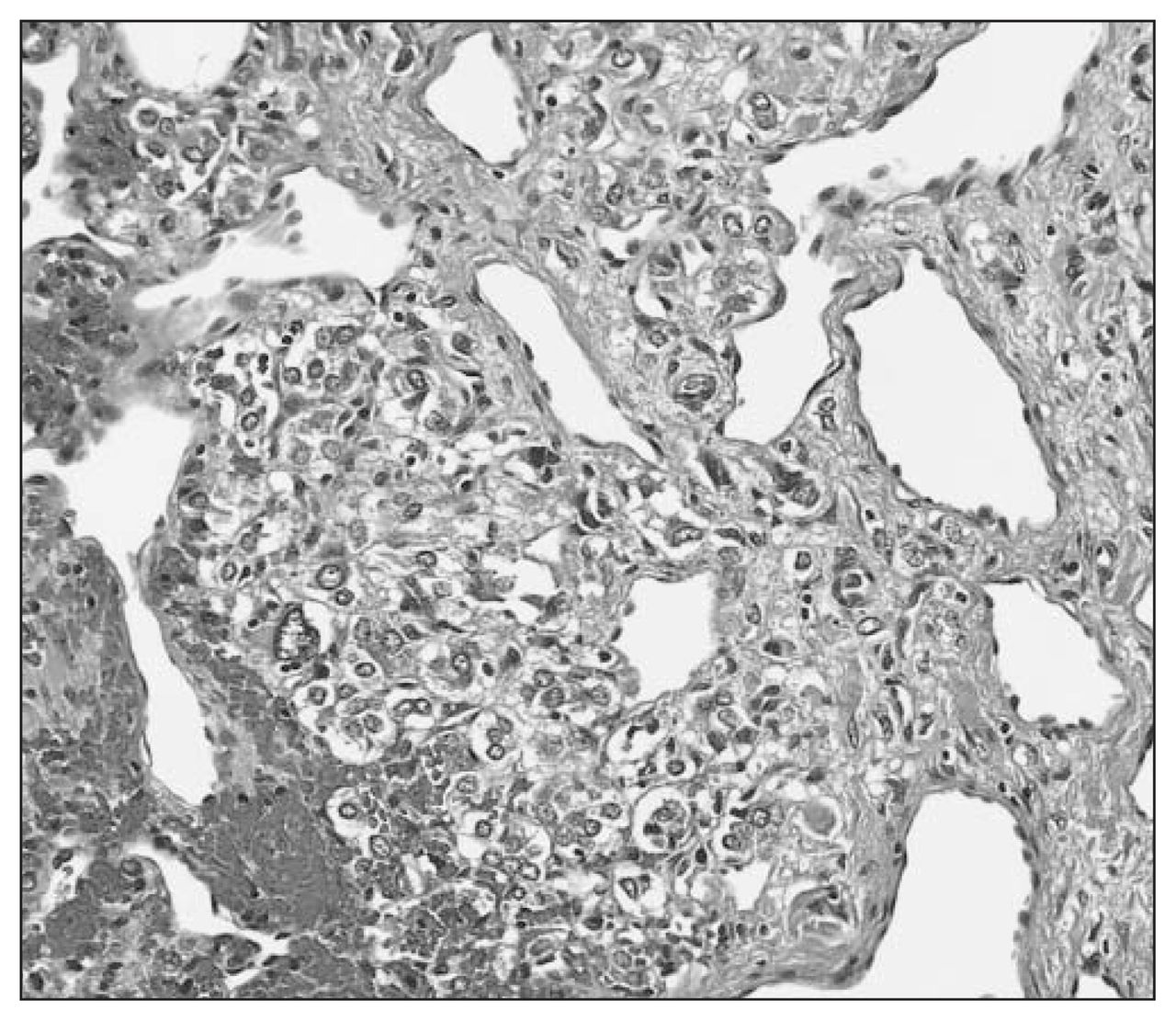
Photomicrograph showing the pheochromocytoma (hematoxylin–eosin, original magnification ×40).
The patient’s postoperative recovery was uncomplicated, and at the 3-month follow-up the urinary catecholamine levels were normal.
Footnotes
Submissions to Surgical Images, soft-tissue section, should be sent to the section editors: Dr. David P. Girvan, Victoria Hospital Corporation, PO Box 5375, Station B, London ON N6A 5A5 or Dr. Nis Schmidt, Department of Surgery, St. Paul’s Hospital, 1081 Burrard St., Vancouver BC V6Z 1Y6.
Competing interests: None declared.
In this issue

Article tools


Related Articles
Cited By...
- No citing articles found.