


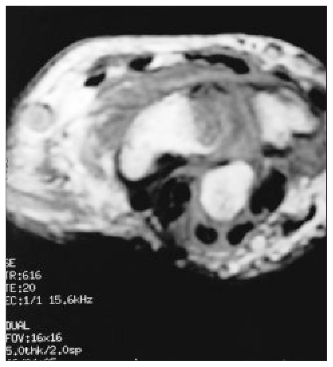
A30-year-old man was referred for magnetic resonance imaging of a painful right wrist. He had been involved in a motorcycle accident several days earlier and had fallen onto his wrist. Fig. 1 is an axial T1-weighted image through the radiocarpal joint. Fig. 2 is a coronal T1-weighted image. Dorsovolar and lateral plain radiographs were subsequently obtained for correlation with the magnetic resonance images and are shown in Figs. 3 and 4.

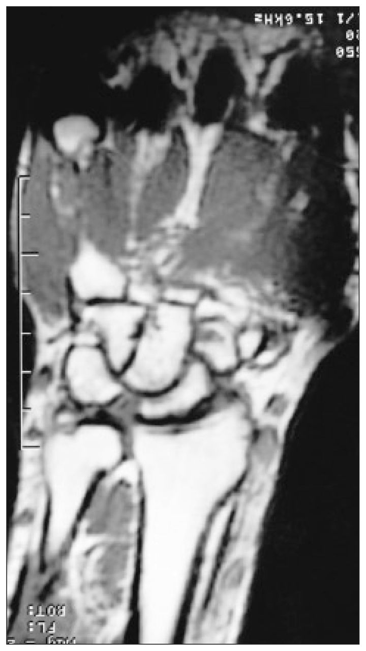
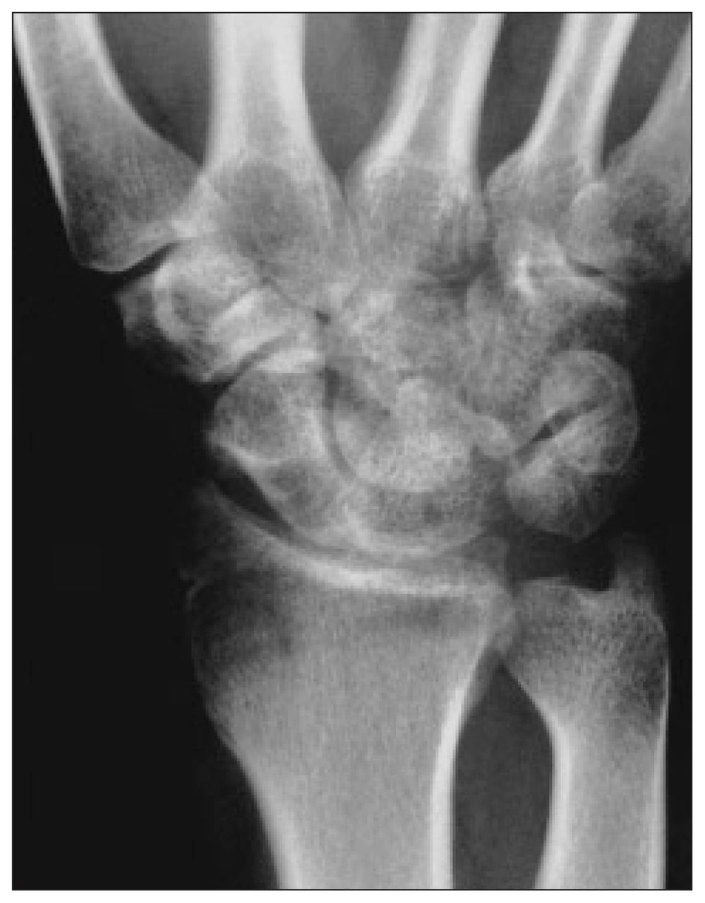
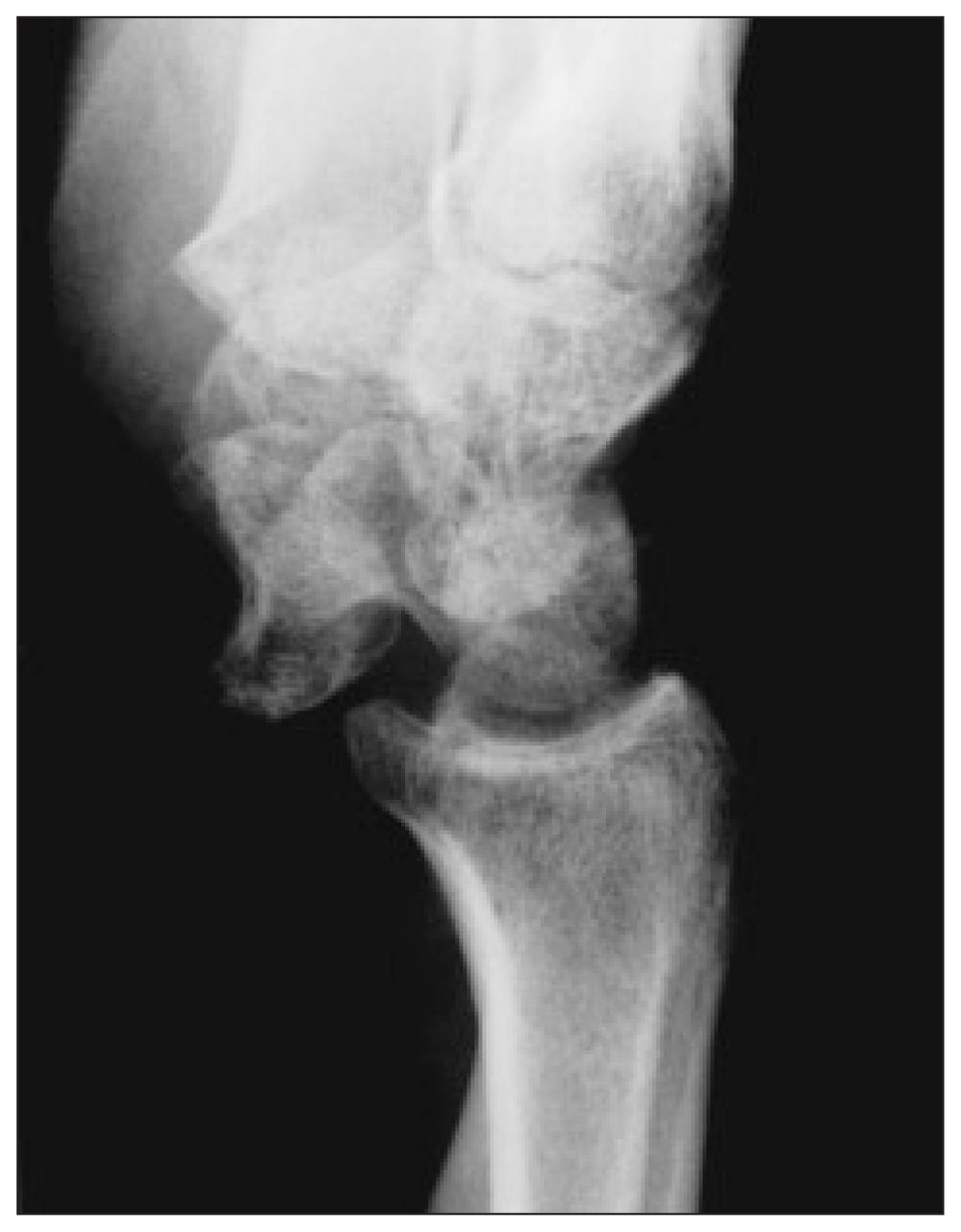
From these images and radiographs can you make the diagnosis?
For the answer and discussion see page 307.
Footnotes
Section Editor: Peter L. Munk, MD
Inquiries about this section should be directed to Dr. Peter L. Munk, Professor, Department of Radiology, Vancouver General Hospital and Health Sciences Centre, 855 West 12th Ave., Vancouver BC V5Z 1M9.
In this issue

Article tools


Related Articles
Cited By...
- No citing articles found.