


A 52-year-old man with a history of cholecystectomy for symptomatic gallstones and hepatitis B liver cirrhosis presented with epigastric discomfort. Ultrasonography and computed tomography showed 2 lesions in the right hepatic lobe. Transarterial chemoembolization had been carried out as the primary treatment at another hospital. He had also taken traditional Chinese herbal products before he was referred to our unit.
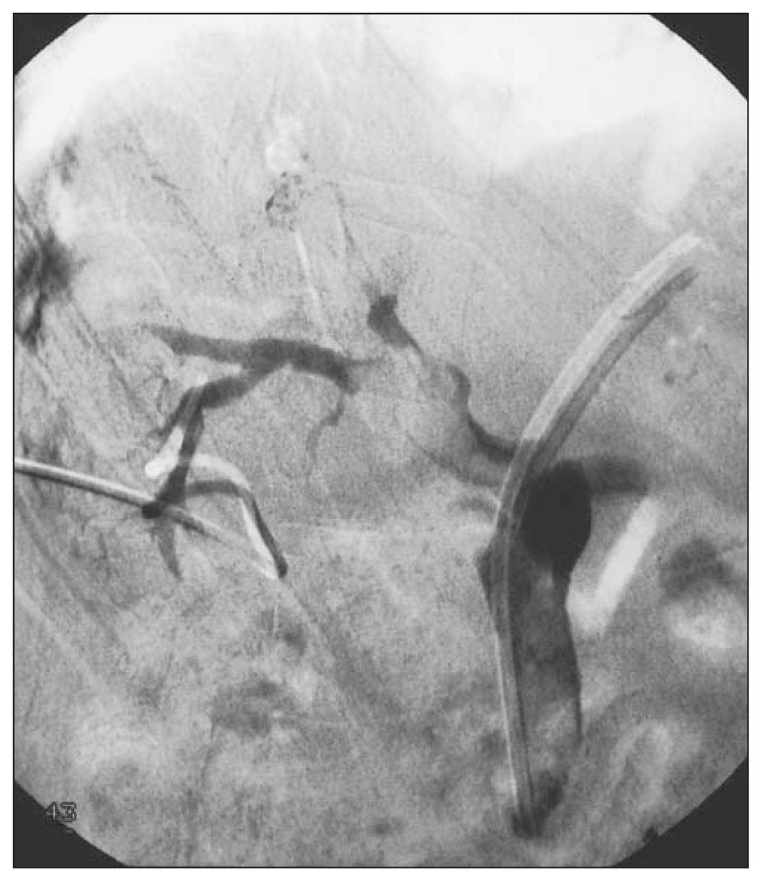
On admission he had an elevated α-fetoprotein level (221 μg/L). Computed tomography (Fig. 1) showed a dilated biliary tree, 2 enhancing lesions in segments 6 and 7 and another lesion in segment 4. He also had obstructive jaundice. The serum bilirubin level was elevated (243 μmol/L) as was the serum alkaline phosphatase (316 U/L). Ultrasonography confirmed dilatation of the intrahepatic ducts and the common hepatic duct, with soft-tissue echoes within the common bile duct. Endoscopic retrograde cholangiopancreatography (Fig. 2 [next page]) was done, and an internal stent was inserted into the left ductal system. The serum bilirubin level showed an initial downward trend to approximately 50 μmol/L but no further decrease toward normal. Percutaneous transhepatic drainage was then done to drain the undrained segments in the right lobe (Fig. 3 [next page]).


What is the diagnosis and what abnormalities are demonstrated on the cholangiograms?
For the diagnosis and a discussion see page 311.
Footnotes
Submissions to Radiology for the Surgeon, soft-tissue section, should be sent to the section editor: Dr. Lawrence A. Stein, Department of Radiology, Royal Victoria Hospital, 687 Pine Ave. W, Montréal QC H3A 1A1; lawrence.stein{at}muhc.mcgill.ca
In this issue

Article tools


Related Articles
Cited By...
- No citing articles found.